
Fig. 3.
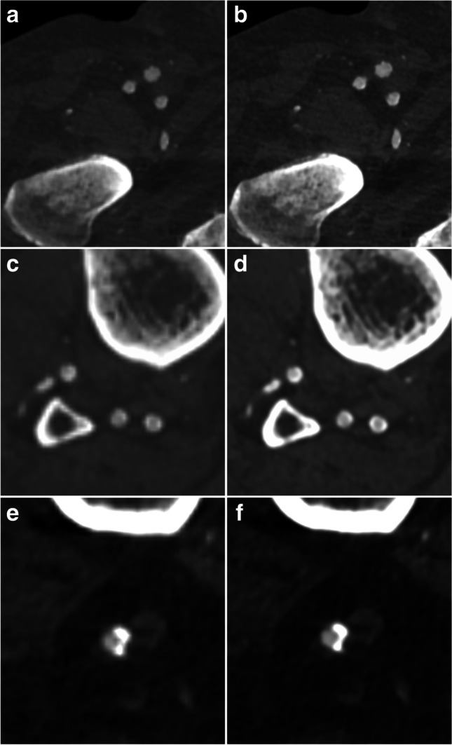
Illustration of the improvement of image quality for the small FOV reconstruction (right column) as compared to the standard reconstruction (left column). Already at the femoral level (a, b) a sharper image quality and better delineation of the vessel wall is noted. The effect is much more pronounced at the calf level (c, d) in the same patient with heavily calcified arteries. In another patient with severe vessel calcifications of the popliteal artery (e, f), the reconstruction with a smaller FOV results in a better delineation of the contrasted vessel lumen against the coarse calcification