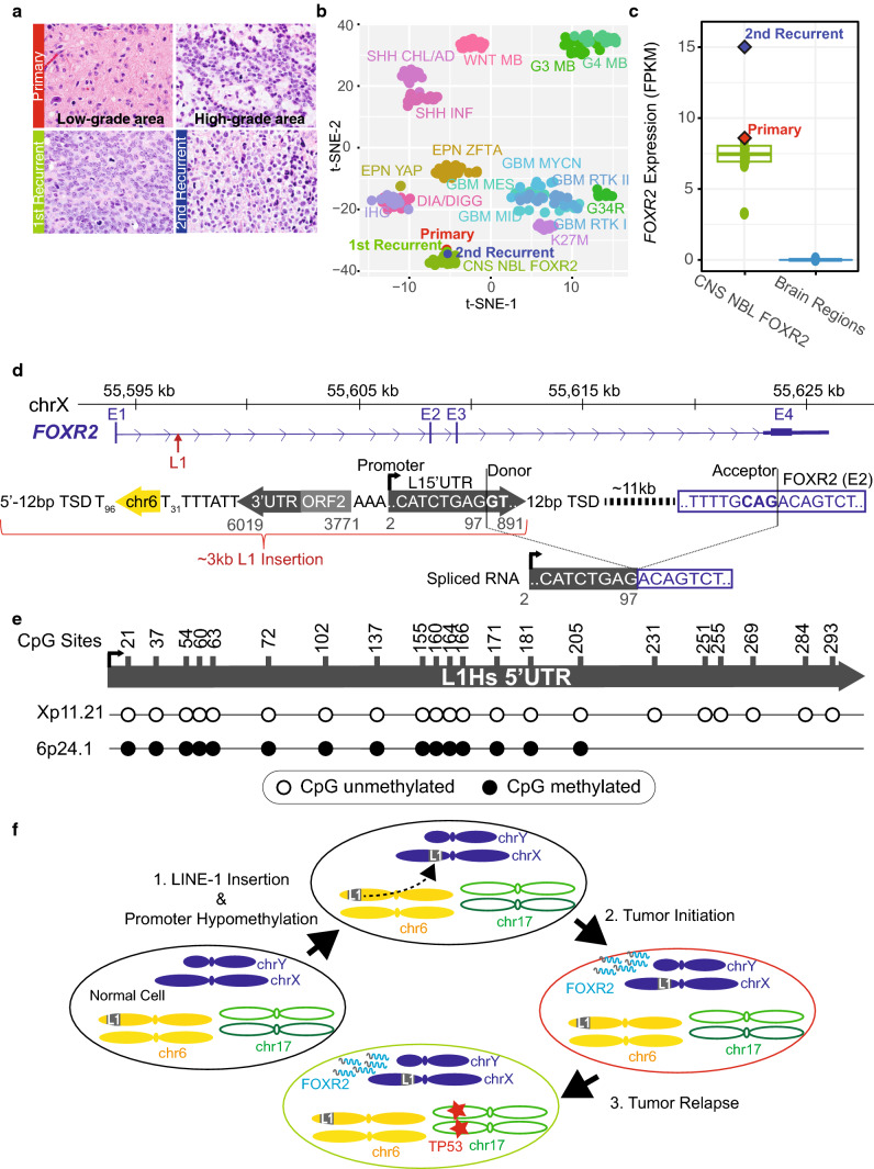
Fig. 1.
Somatic L1 promoter donation drives oncogenic FOXR2 overexpression. a Histology of serial tumor samples. b t-SNE plot comparing genomic DNA methylation profiles of the primary and recurrent tumors to 249 pediatric CNS tumors of 17 types (see Supplementary text, online resource for additional abbreviations). c FOXR2 expression of CNS NBL FOXR2 overlayed by the primary and 2nd recurrent tumors as compared to non-diseased multiple brain regions profiled by GTeX. d Top, schematic of PacBio sequenced L1 insertion with a chr6 transduction sequence (yellow arrow) and flanked by target-site duplication sequence (TSD). Gray numbers match nucleotides of L1.3 consensus sequence. Below, observed tumor transcripts involve L1 splice donor to canonical FOXR2 splice acceptor. e Methylation status of CpG sites (nucleotide numbers) in the L1 5’UTR at the retrotransposed FOXR2 locus (Xp11.21) and the source element (6p24.1) observed in over 90% of bisulfite sequencing reads. f Model of oncogenic activation with chr6 L1 source element (gray ‘L1’ box) insertion in chrX upstream of FOXR2, inducing oncogenic overexpression of FOXR2 (gray and blue squiggles), driving the primary tumor. Recurrent tumor formed, acquiring a TP53 R175H variant (red star)

