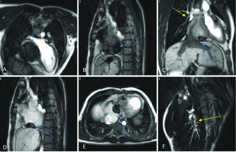
Fig. 24.

Horizontal long-axis (A), sagittal (B, D), coronal (C), axial (E) and right pulmonary artery cross-cut views (F) of a cardiovascular MRI study in an atriopulmonary Fontan circulation performed for congenital tricuspid atresia. There are filling defects in keeping with thrombi in the right atrium (blue arrows) and in the right pulmonary artery (yellow arrows) on inversion recovery early-phase gadolinium sequences. Note that thrombus appears as very hypointense. Re-used with permission from Fontan circulation in an adult: A guide for the radiologist. Arzanauskaite M, Nyktari E, Voges I. ESTI-ESCR 2018/P-0103
