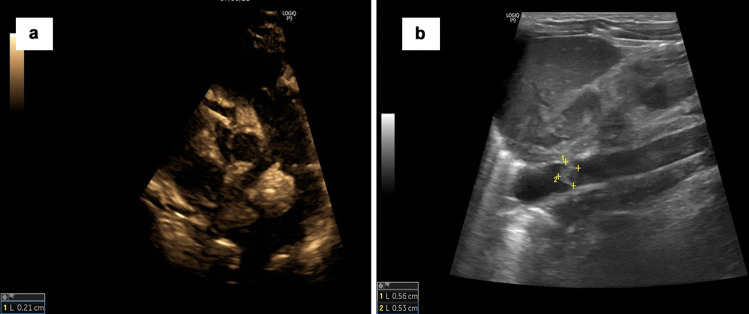
Fig. 1.
a Echocardiography image of the first case showing dilated right coronary artery (RCA) measuring 2.1 mm (+3.58 z score); the baby also had dilated left main coronary artery (LMCA) of 2.1 mm (+2.04 z score) and left anterior descending artery (LAD) of 2 mm (+4.49 z score) (not shown in the figure). b Ultrasound image of the second case showing 5.6 mm × 5.3 mm thrombus in abdominal aorta just above the left renal artery origin

