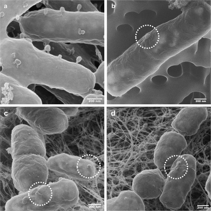
Fig. 1. Helium ion microscopy (HIM) visualization of T4 phage adsorption to E. coli host and non-host P. putida KT2440 cells.
a, b visualize cells from phage adsorption experiments (cf. materials and methods) reflecting tail-mediated adsorption to E. coli host cells (a) and capsid-driven adsorption to non-host P. putida KT2440 (b). c, d visualize tail- (c) and capsid-driven (d) phage adsorption to biofilm cells growing on agar patch B on day 2 in experiments evaluating population effect of T4 co-transport with P. putida KT2440 (cf. Fig. 3).

