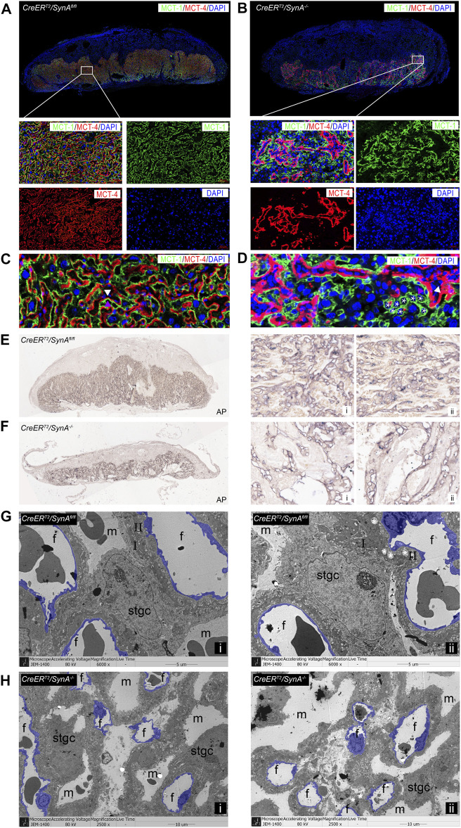
FIGURE 2.
Abnormal formation of the labyrinth layer in the CreER T2 /SynA −/− placenta. (A,B) Anti-MCT1 and anti-MCT4 immunofluorescence staining of the CreER T2 /SynA fl/fl and CreER T2 /SynA −/− placentas at E17.5 d. Anti-MCT1 and anti-MCT4 are expressed in the labyrinth, which showed a reduced labyrinth area in the CreER T2 /SynA −/− placenta. (C,D) Unfused trophoblast cells gathered in the labyrinth layer, surrounding the morphologically abnormal STB-II, indicating that STB-I was disrupted. Moreover, the STB-II was reduced after Syncytin-A disruption. (E,F) AP staining of CreER T2 /SynA fl/fl and CreER T2 /SynA −/− placentas at E17.5 d. Overall figure (4×), partial figure (20×). (G,H) Transmission electron microscopy observation of endothelial cells (purple) in placental tissues at E17.5 d. (G) (6,000×), (H) (2,500×, for better insight on vascularization in CreER T2 /SynA −/− placentas). m: maternal lacuna; f: fetal vessel; I: STB-I; II: STB-II; *: lipid inclusion.

