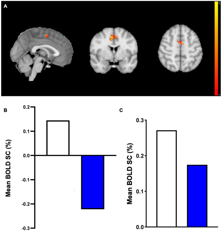
FIGURE 5.
Anterior cingulate cortex region of interest fMRI faces task results. A region of interest activation for the anterior cingulate cortex (ACC) in response to mean effect of task in placebo vs. prucalopride group. Sagittal, coronal, and axial images (shown at MNI location 45,63,62) depicting significantly increased activation in the placebo group for the mean > fixation contrast [placebo > prucalopride, Z = 4.39, p < 0.0002, peak voxel location: x = 6, y = 0, z = 50, cluster size = 164 voxels]. Images thresholded at z > 3.1, p < 0.05 corrected. Red to yellow colours identify increases in brain activation (scale Z = 3.1–4.3). ACC Mask = Harvard Oxford atlas. (B) Group mean of BOLD percentage signal change (SC) extracted from the cluster in (A). White = placebo, blue = prucalopride. (C) Group mean of BOLD percentage signal change (SC) extracted from functional anterior cingulate cortex (ACC) mask in response to mean effect of task. A functional ROI (SVC) mask was created for the ACC for mean > fixation by multiplying mean activation for all participants by the anatomical mask. White = placebo, blue = prucalopride.

