Figure 1.
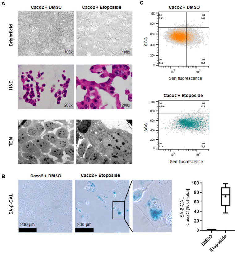
Induction of cellular senescence in vitro by Etoposide treatment. (A) Morphological appearance of Etoposide (Eto)-treated and control (DMSO) Caco2 cells using brightfield microscopy (upper panel), normal H&E-stained sections of cytoblocks (middle panel) and TEM (lower panel). Senescent cells adopt a characteristic “fried egg” morphology, including an enlarged shape and nucleus. Cytoplasmatic vacuolation is apparent. (B) Increased senescence SA-β-GAL staining indicates senescence induction. SA-β-GAL-positive cells were counted after DMSO and Etoposide-treatment. (C) FACS analysis confirms senescence induction in Etoposide-treated cells.

