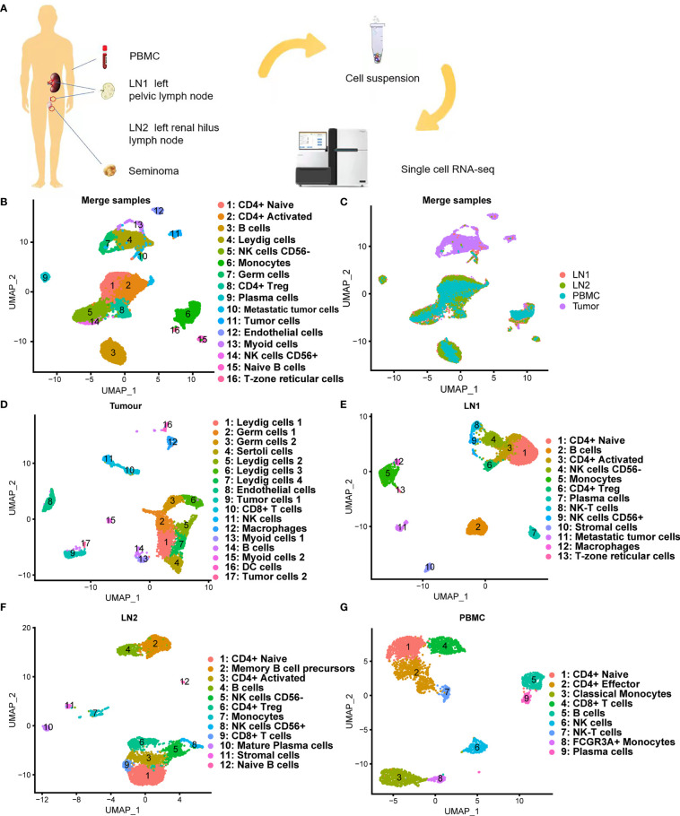
Figure 2.
Single-cell transcriptomic atlas of testicular seminoma sample. (A) Schematic design of the overall design. (B, C) Single-cell transcriptomic map of testicular tumor (Tumor), left pelvic lymph node (LN1), left renal hilus lymph node (LN2) and peripheral blood mononuclear cells (PBMC), with 16 distinct cell types. (D) UMAP plot representation of testicular seminoma (Tumor) with 17 distinct cell types. (E) UMAP plot representation of left pelvic lymph node (LN1) with 13 distinct cell types. (F) UMAP plot representation of left renal hilus lymph node (LN2) with 12 distinct cell types. (G) UMAP plot representation of PBMCs with 9 distinct cell types.

