Figure 3.
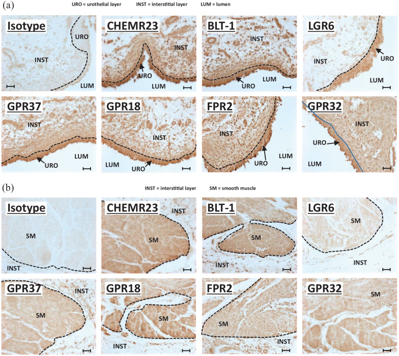
SPM receptors are expressed in human bladder: (a) micrographs focused on the urothelia and underlying interstitial cells, (b) micrographs focusing on the bladder smooth muscle. All panels in (a) and (b) show immunohistochemistry performed on 5-µm sections of formalin-fixed, paraffin-embedded human bladders purchased from Zyagen Inc. (San Diego, CA). The primary antibody target is indicated on each panel and all were rabbit antibodies (see Table 1). The first panel in both (a) and (b) is an isotype control using a rabbit IgG isotype control from Novus Biologicals (Centennial, CO) (Table 1). All parameters for the slides used in this figure (incubation times, HRP product development times, etc.) were identical between all samples and for each antibody and all slides specifically used in this figure were stained on the same day. Micrographs were taken with identical settings (exposure times, light intensity etc.). Scale bars = 50 µm. The various layers (urothelial, interstitial, and smooth muscle) are delineated with dash lines and labeled on the micrographs.

