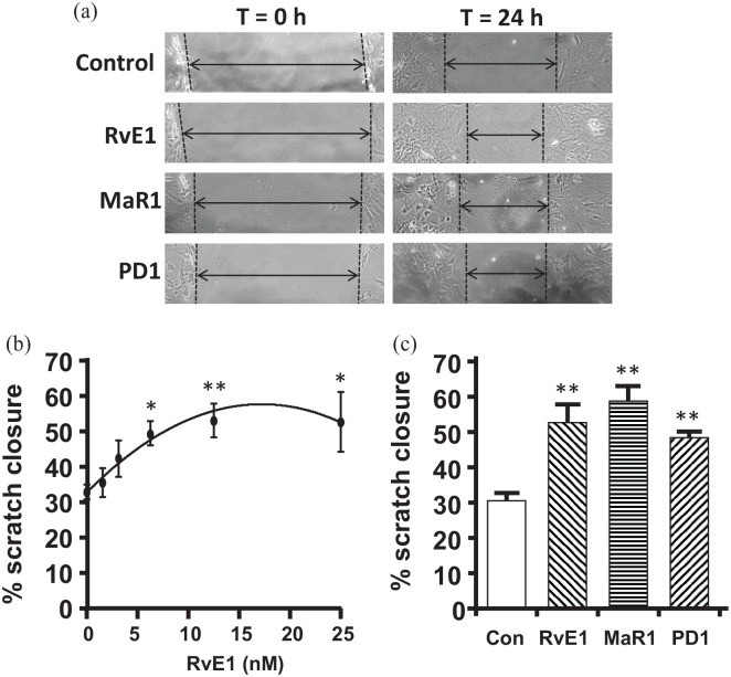
Figure 4.
RvE1, MaR1, and PD1 enhance wound closure in the in vitro scratch assay: (a) representative micrographs of the scratch wound in the monolayer of urothelia before (t = 0 h) and after (t = 24 h) treatment with the indicated SPM or an equivalent volume of PBS, as described in the methods section. (b) Dose response of RvE1. As described in the Methods section, mouse urothelia were placed in culture and allowed to form a monolayer. A scratch was then made and photographed. RvE1 was added at the indicated doses and, after an additional 18 h, the scratch photographed at the identical spot. The width of the scratch was determined at both time points and the % of the scratch closure determined. Results are the mean ± SEM. n = 10, 5, 6, 5, 5, 5, respectively. *p < 0.05, **p < 0.01, ***p < 0.001 by ANOVA and Dunnett’s post hoc test comparing all to 0 nM control. (c) Effects of RvE1, MaR1, and PD1 on scratch closure. The scratch assay was performed as described and cultures were treated with either vehicle or 12.5 nM of RvE1, MaR1, or PD1. The RvE1 value shown is the same data used in (a) but is included in (b) for ease of comparison with the other SPMs. Results are the mean ± SEM. n = 10, 5, 3, 3, respectively. **p < 0.01, ***p < 0.001 by ANOVA and Dunnett’s post hoc test comparing all to control.

