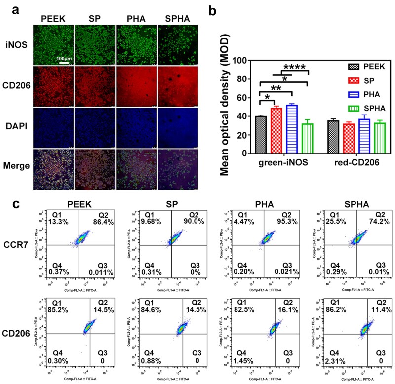
Figure 5.
Immunofluorescence staining images of RAW264.7 cultured on samples for 4 days (a) and corresponding quantitative analysis (b); iNOS (green) was selected as a marker for M1 macrophages, CD206 (red) was selected as a marker for M2 macrophages and the nuclei was stained by DAPI (blue). Flow cytometry analysis of RAW264.7 cultured on samples after 4 days (c).

