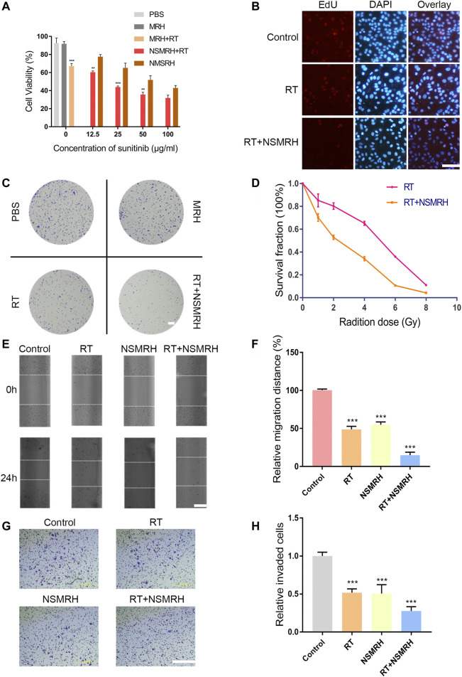
FIGURE 3.
In vitro functions of NSMRH in MM-231 cells. (A) Effects of different conditions on MM-231 cells viability at 48 h. (B) Edu staining of MM-231 cells, red fluorescence represents proliferating cells, scale bars: 100 μm. (C) A clonogenic assay was performed. Colonies larger than 50 cells were counted, and phase-contrast images were taken, scale bar: 2.5 mm. (D) Survival curves of MM-231 cells exposed to increased irradiation. (E) Effect of NSMRH on MM-231 cells migration. Representative images showing tumor cells migration, scale bars: 100 μm. (F) The figure shows quantitative analysis of a wound healing assay, with triplicate measurements of three independent experiments. (G) Photomicrographs illustrate representative fields of invaded cells, scale bars: 100 μm. (H) The relative number of invaded cells was calculated, and the data are presented in a histogram from three independent experiments.

