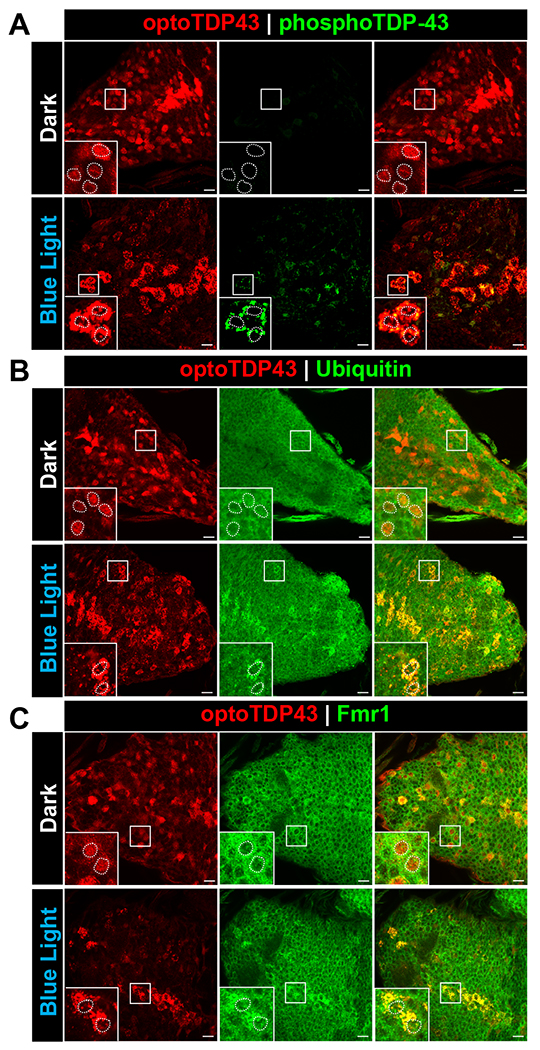
Fig. 2. optoTDP43 cytoplasmic aggregates colocalize with key biochemical markers of TDP-43 proteinopathy.

ok371-GAL4;UAS-optoTDP43 larvae were raised at 25 °C and kept under blue light or kept in the dark for 24 hours prior to processing for processing for immunofluorescence. A. Representative images of hyperphosphorylated cytoplasmic optoTDP43 aggregates in ok371-GAL4;UAS-optoTDP43 larvae motor neurons following 24 hours of blue light illumination. These assemblies were not observed in larvae kept in darkness. B. Representative images of ubiquitinated cytoplasmic optoTDP43 aggregates in ok371-GAL4;UAS-optoTDP43 larvae motor neurons following 24 hours of blue light illumination. These assemblies were not observed in larvae kept in darkness. C. Cytoplasmic optoTDP43 colocalize with Fmr1, a marker of Drosophila RNA granules, in ok371-GAL4;UAS-optoTDP43 larvae motor neurons following 24 hours of blue light illumination. n = 5 larvae per staining. Scale bar = 10 um.
