
Fig. 2. Cell fates of blastomeres tagged at the 2-cell stage of development with DiI-CM.
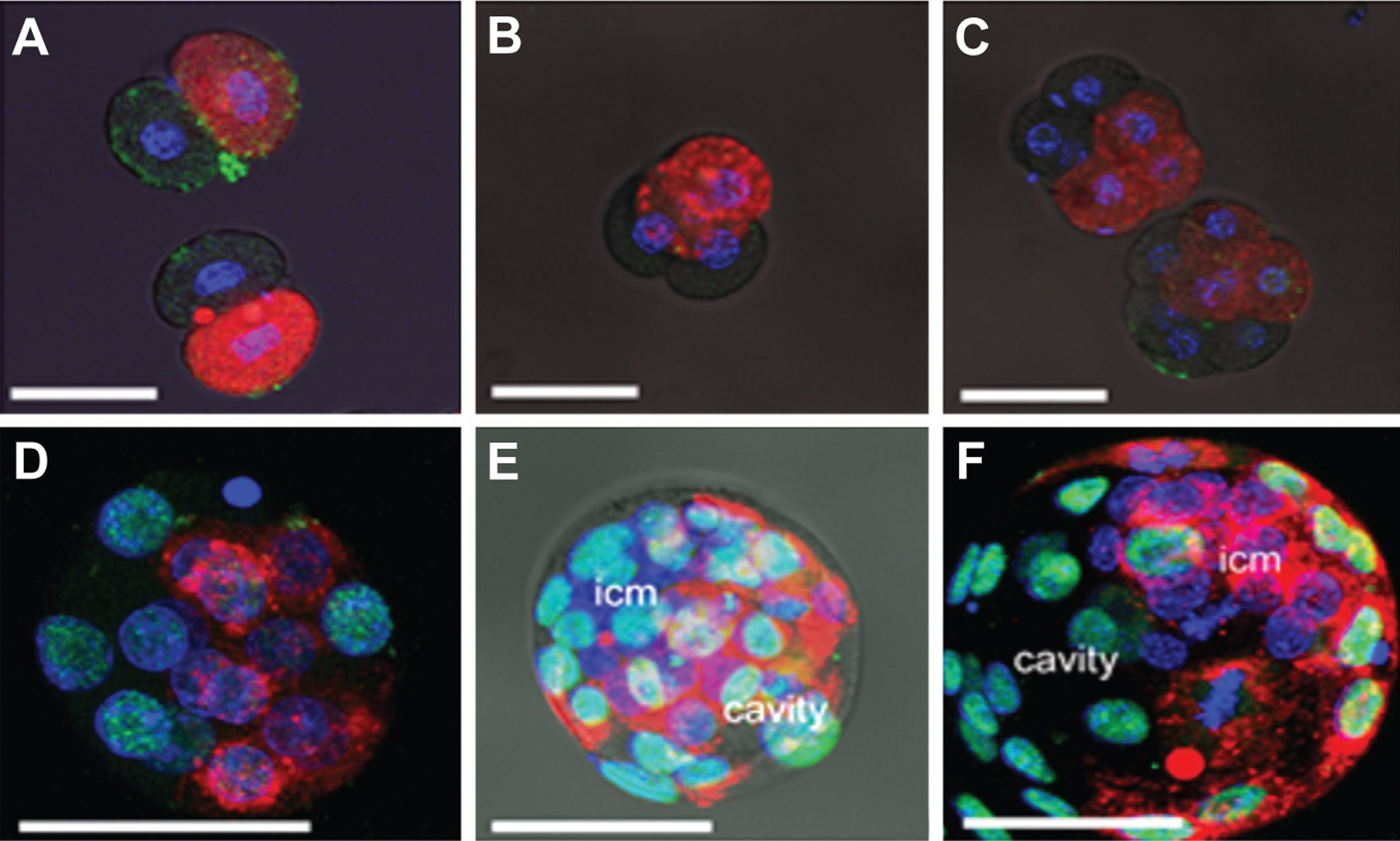
After tagging one randomly chosen blastomere with the membrane dye, DiI-CM (red) at the 2-cell stage of development, embryos were allowed to proceed in their development through various intermediary stages until they reached blastocyst. Each embryo was counter stained with DAPI (blue) and anti-Cdx2 antibody (green). (A) 2-cell stage embryos; (B) 4-cell stage; (C) 8-cell stage; (D) morula. In blastocysts, labeled cell progeny were frequently concentrated towards either the abembryonic pole [(E) red cells located opposite the ICM] or the embryonic pole [(F) red cells associated with the region occupied by the ICM and polar trophectoderm]. Supplementary Fig. S1 A–F provides the original confocal stacks for Fig. 2 A–F, and also for a blastocyst with a typically random distribution of DiI (Supplementary Fig. S1G) in which there was no preferential localization to either the embryonic or abembryonic poles.