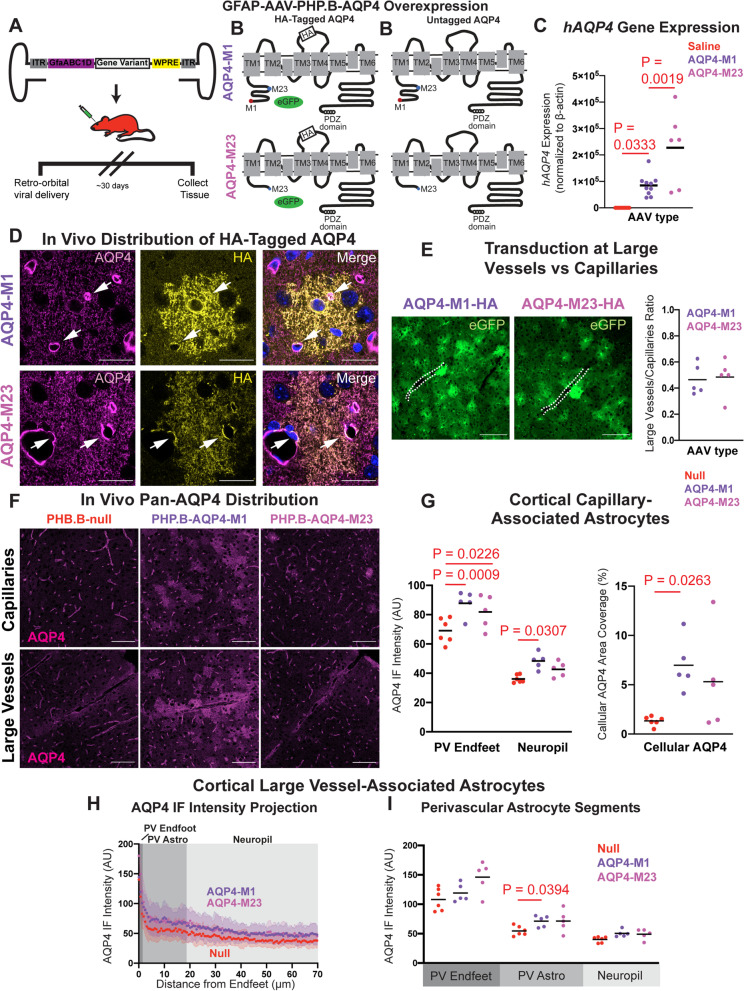
Fig. 3.
Viral AQP4-M1 overexpression increases cellular AQP4 localization in mouse cortex. A Schematic outline of the viral approach to overexpress AQP4-M1 or -M23 isoforms under the astrocyte-specific GfaABC1D promoter in vivo. B AAVPHP constructs were generated to drive expression of untagged human AQP4 (hAQP4)-M1 and -M23 isoforms (right), or co-expression of HA-tagged hAQP4-M1 and -M23 with enhanced green fluorescent protein (eGFP) reporter. C Thirty days after retro-orbital viral delivery, qPCR showed that untagged hAQP4-M1 and hAQP4-M23 were robustly expressed in the mouse cortex (P = 0.0333, P = 0.0019; one-way ANOVA with Tukey’s post hoc correction). D Thirty days after iv viral delivery, HA-tagged AQP4-M1 exhibited no specific perivascular (white arrows) localization, distributing over all fine processes (top) while HA-tagged AQP4-M23 localized to both perivascular endfeet and fine astroglial processes (bottom). Scale bar: 20 μm. E Evaluation of eGFP reporter expression showed that viruses driving AQP4-M1 (top image) and AQP4-M23 (bottom image) efficiently transduced astrocytes surrounding both large cortical vessels (white dash lines) and capillaries, and that the transduction ratios along large vessels and capillaries were comparable (panel at right). Scale bar: 100 μm. F Thirty days after delivery of virus driving untagged AQP4-M1 and -M23, AQP4 IF was evaluated with a pan-AQP4 antibody in capillaries (top) and large cortical vessels (bottom). AQP4-M1 overexpression resulted in a diffuse pattern of AQP4 labeling in the fine processes of astrocytes throughout the cortex while AQP4-M23 overexpression did not result in marked cellular labeling. Scale bar: 100 μm. G In cortical capillary-associated astrocytes, overexpression of AQP4-M1 isoform significantly increased the AQP4 IF intensity at both PV Endfeet and in the surrounding non-perivascular neuropil (Left, P = 0.0009, P = 0.0307, respectively. 2-way ANOVA with Tukey’s post hoc test), as well as the cellular AQP4 area coverage (Right, P = 0.0263, 1-way ANOVA with Tukey’s post hoc test). On the contrary, overexpression of AQP4-M23 isoform only increased the AQP4 IF intensity at the PV Endfeet (left; P = 0.0226, 2-way ANOVA with Tukey’s post hoc test). H,I Cross-sectional analysis of AQP4 IF surrounding large cortical vessels showed that compared to control (null) virus (30 vessels from 6 animals), AQP4-M1 (31 vessels from 5 animals) overexpression significantly increased AQP4 IF in PV Astrocytes segments (P = 0.0394, 2-way ANOVA with Tukey’s post hoc test) while AQP4-M23 (27 vessels from 5 animals) did not

