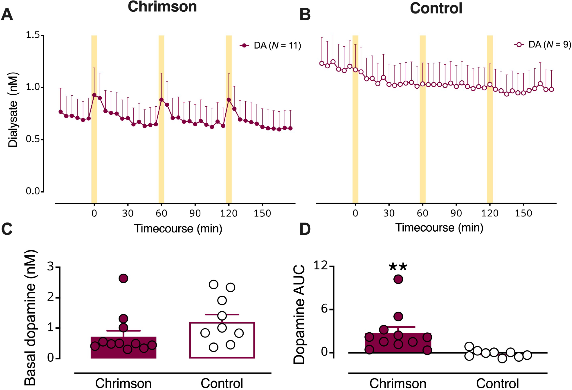
Figure 2: Optical stimulation of dopaminergic cell bodies produces dopamine release in striatal terminal regions.

(A) Dialysate dopamine levels were increased in response to optical stimulation in mice expressing Chrimson (N=11) (B) but not in control mice (N=9). The yellow bars indicate optical stimulations (10 Hz @ 10 mw/mm2, 50 ms pulses, for 5 min). C. Basal dopamine levels in mice transfected with Chrimson relative to control mice. D. Dopamine overflow, quantified by area under the curve, was increased in Chrimson expressing but not control mice. Data are means ± SEMs. **P<0.01.
