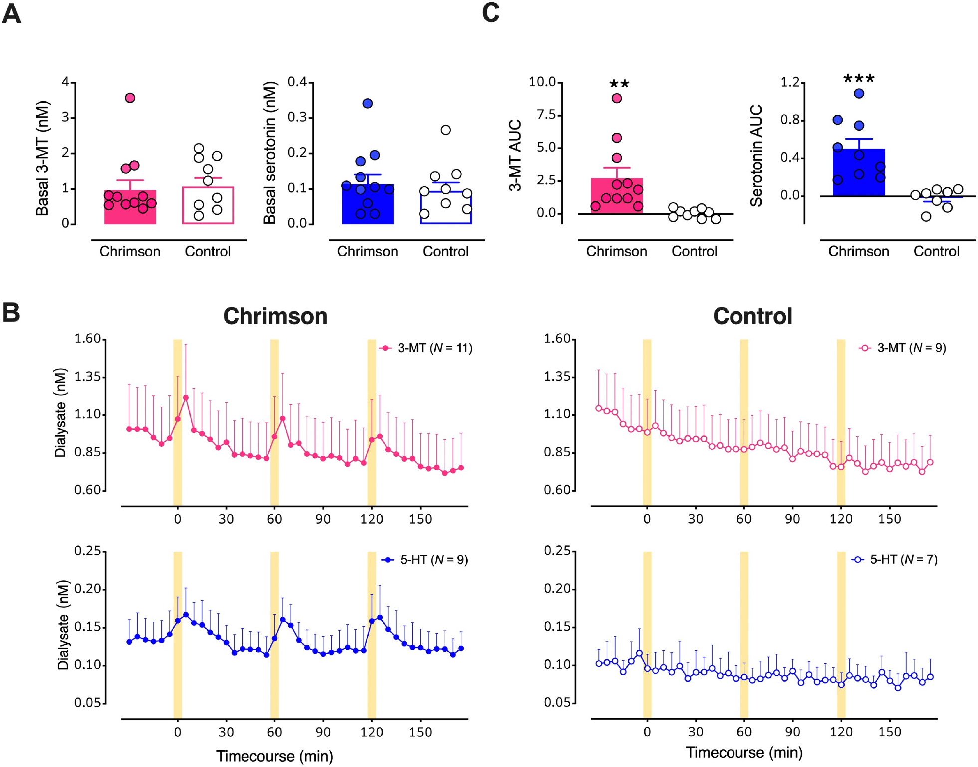
Figure 4: Optical stimulation of midbrain dopamine neurons evokes overflow of 3-methyltyramine (3-MT) and serotonin in dorsal striatum (dSTR).

A. Basal dialysate levels of 3-MT (left, pink) and serotonin (right, blue) in mice with vs. without Chrimson transfection. B. Time course of stimulated 3-MT (pink) and serotonin (blue) in mice transfected with Chrimson (left) compared to mice transfected with a control protein (right). Yellow bars indicate 5-min optical stimulations. C. Comparisons of areas under the curve (AUC) for the overflow of 3-MT or serotonin produced by optical stimulation of dopamine neurons expressing Chrimson with respect to control mice. Data are means ± SEMs. **P<0.01, ***P<0.001. In two mice per group, data for serotonin were below the detectable limit.
