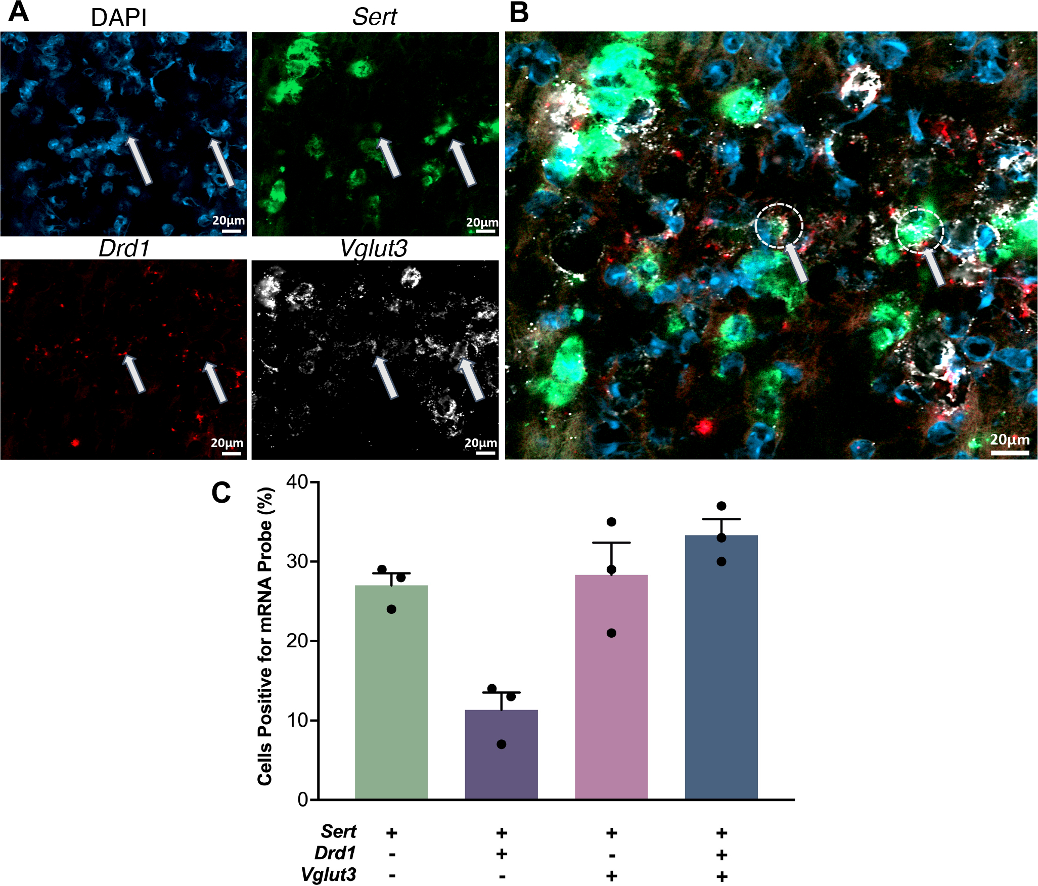
Figure 5: Co-localization of serotonin transporter (Sert), D1 dopamine receptor (Drd1), and vesicular glutamate transporter 3 (Vglut3) mRNA in the dorsal raphe nucleus.

A. Cell nuclei were stained with DAPI (top left, blue). Antisense probes to localize Sert (top right, green), Drd1 (bottom left, red), and Vglut3 (bottom right, white) mRNA were visualized. Puncta for each mRNA were colocalized in some nuclei but did not necessarily overlap. B. Overlay of images in A. Arrows indicate examples of the three mRNAs colocalized in the same nuclei. C. Relative quantification of cells containing Sert, Drd1, and Vglut3 mRNA with respect to the total number of Sert expressing cell bodies. (SEMs are for n=3 z-stack planes in a single mouse. A total of 248 cells were counted).
