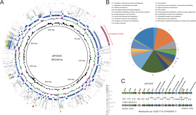
FIG 5.
Structure and recombination analysis of the plasmid pBY2G20. (A) Circular comparison between the plasmid pBY2G20 and other reported similar genomes. Rings represent the following features labeled from inside to outside: ring 1, GC content; ring 2, GC-skew; rings 3 and 4, blue arrows correspond to plus-strand CDS and minus-stand CDS; rings 5 to 8, circular comparison of Metabacillus sp. KUDC1714, Priestia megaterium S2, Bacillus simplex SH-B26, Brevibacterium frigoritolerans zb201705, Brevibacterium sp. PAMC23299, and Priestia flexa 1-2-1, respectively; ring 9, blocks correspond to ISs. (B) Distribution of COG categories for plasmid genes. (C) Genetic organization and comparison of the homologous region between pBY2G20 and the chromosome of Metabacillus sp. KUDC1714. Color coding for genes is based on COG categories. The percent amino acid identities of each homolog are shown.

