
Figure 1.
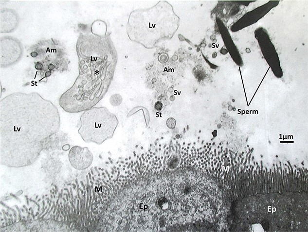
An electron micrograph of mouse spermatozoa in the lumen of oviduct’s isthmus about the time of ovulation after natural mating. Note the presence of many globular and vesicular materials in the isthmus lumen. Am, amorphous material; EP, mucus epithelial cell of isthmus; Lv, large vesicle; M, microvilli of mucus epithelial cell; St, cross section of sperm tail; and Sv, small vesicle. This electron micrograph was prepared by Dr. Kiyotaka Toshimori after perfusion fixation of mouse oviduct.