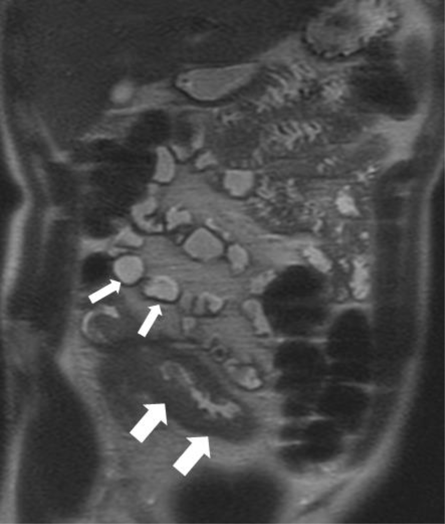
FIGURE 1:

Coronal T2 weighted image in a patient with history of Crohn’s disease demonstrating marked thickening of the terminal ileum (thick white arrows). Normal, unaffected loops of intestine with normal wall thickness (thin white arrows) can also be seen.
