FIGURE 2A:

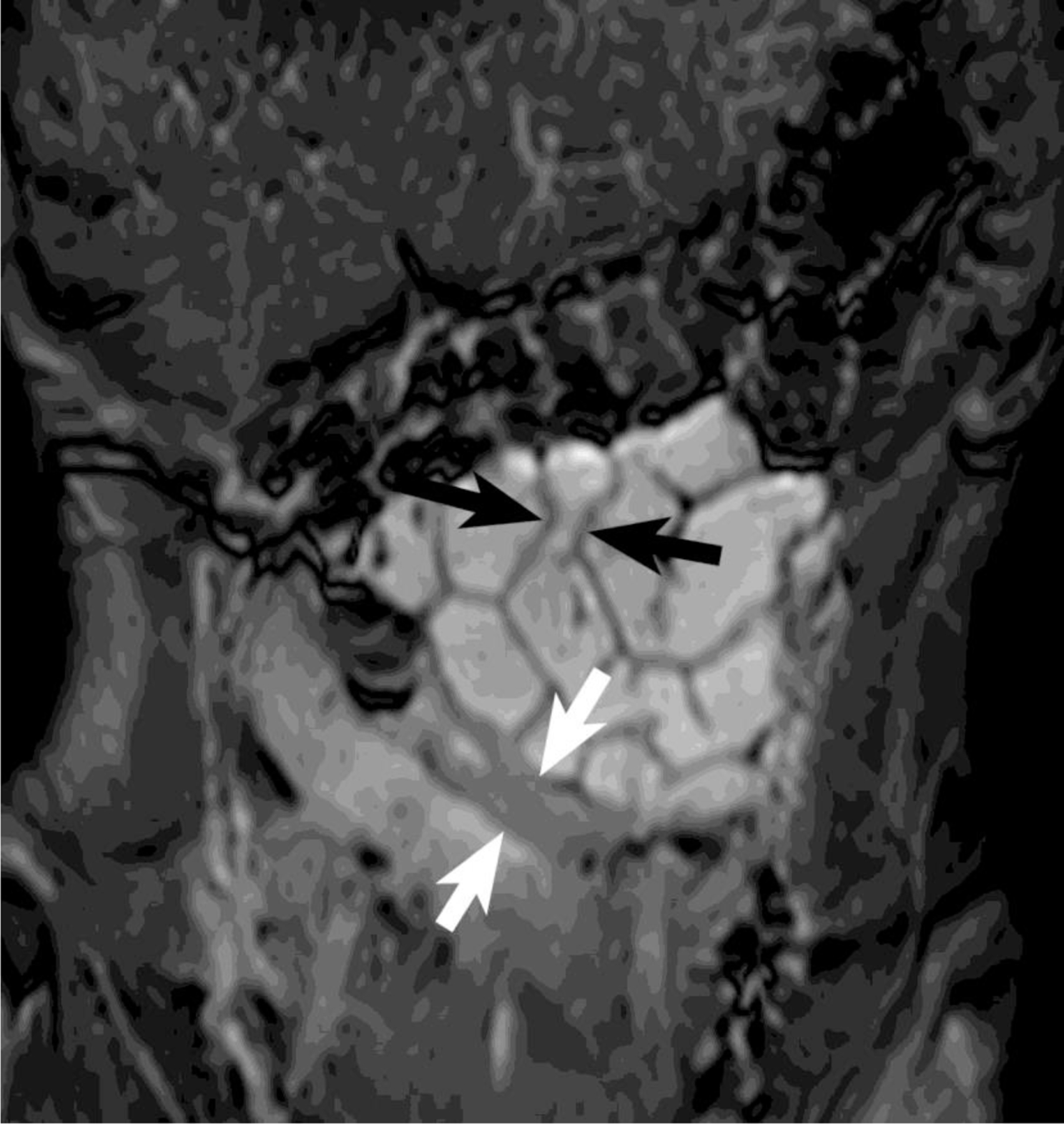
Coronal SSFP image from an MRE in a 15 year old patient with suspected Crohn’s disease. An area of narrowing is seen in the proximal jejunum (black arrows) and more distally in the proximal ileum (white arrows).

Coronal SSFP image from an MRE in a 15 year old patient with suspected Crohn’s disease. An area of narrowing is seen in the proximal jejunum (black arrows) and more distally in the proximal ileum (white arrows).