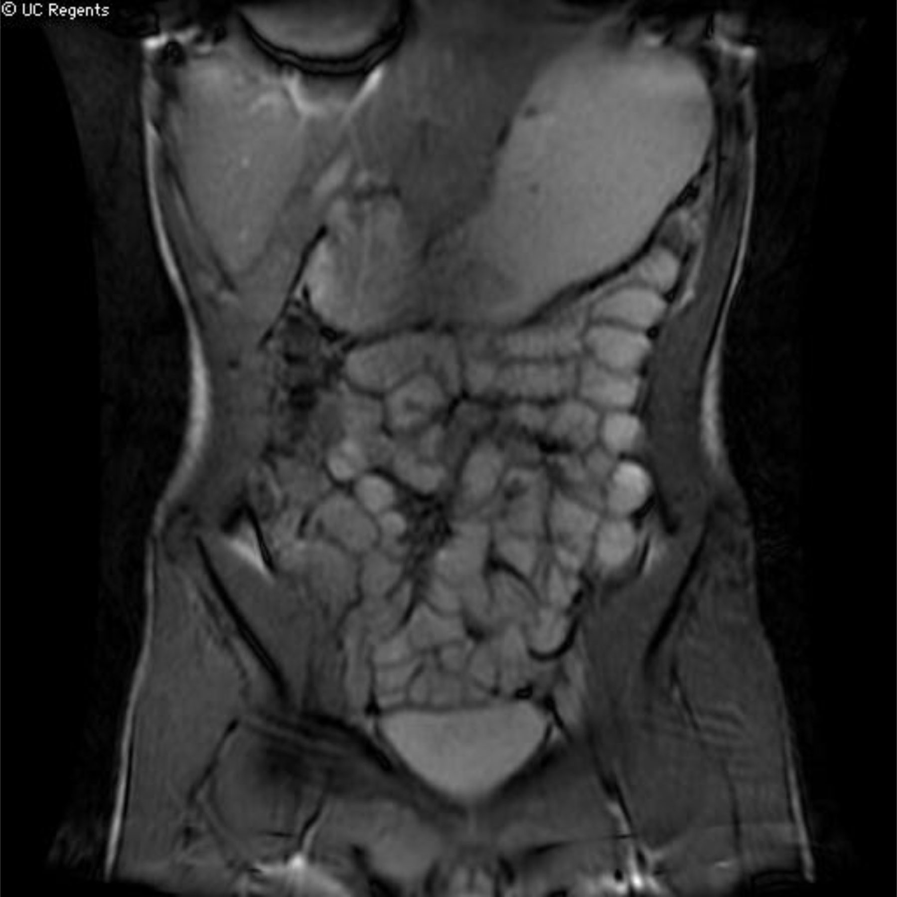
FIGURE 5.

[STILL IMAGE PLACEHOLDER FOR CINE]: Coronal cine SSFP sequence from and MRE performed in a teenage patient with clinically suspected Crohn’s disease. Right lateral decubitus positioning prior to scanning combined with the use of cine SSFP imaging allows for improved evaluation of jejunal loops (white arrows).
