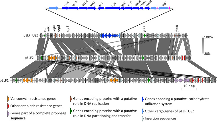
FIG 4.
Genetic structure of pELF_USZ. Adapted and expanded from Fig. 3 of Hashimoto et al. (37). Visualization of the multiple alignment of the three plasmid sequences. Regions shared between sequences are connected by vertical blocks. A minimum blast length of 500 bp and minimum identity of 80% was considered. Color gradient reflects percent identity. Coding sequences are shown with arrows, and their color reflects the genes’ functions. An intact phage identified on pELF1 is highlighted in purple, and the cargo genes of pELF_USZ are highlighted in blue. This 14-kb stretch is amplified for a clearer visualization. The 11 consecutive genes encoding a carbohydrate utilization system are in dark blue, and two tRNA coding genes are in magenta.

