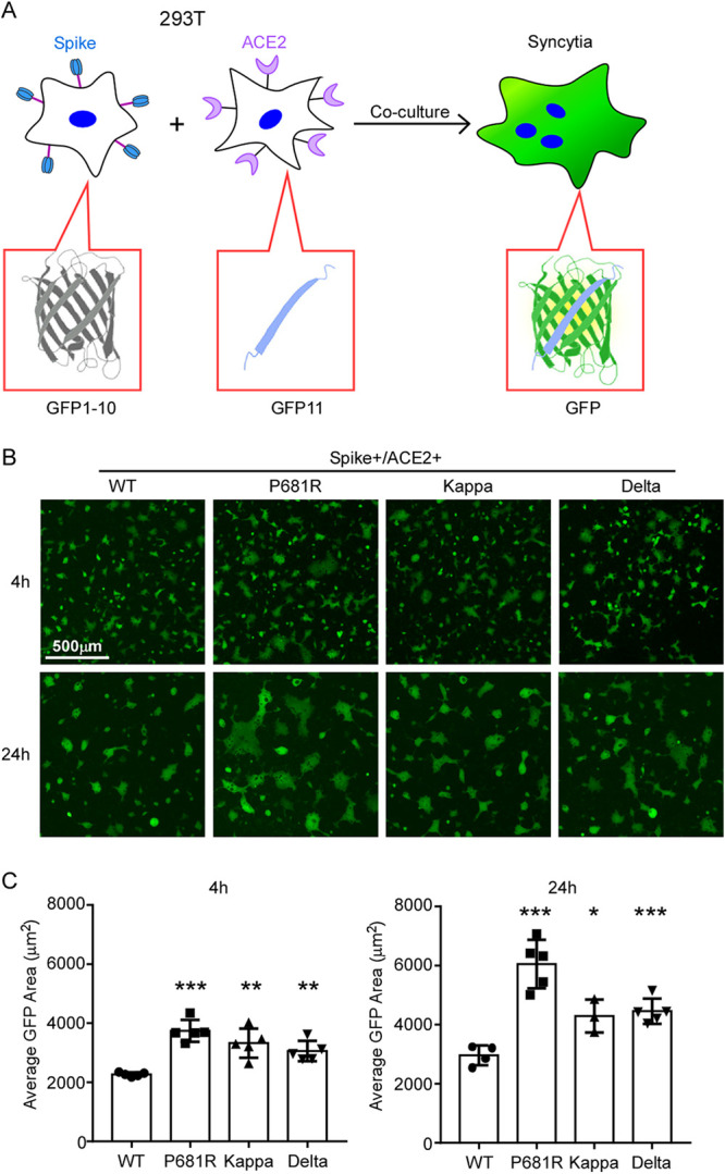
FIG 2.

SARS‐CoV‐2 variant spike protein-mediated syncytium formation. (A) Schematic representation of syncytium formation between the donor cells expressing spike protein and split GFP and the acceptor cells expressing ACE2 receptor and split GFP. The images were taken at 4 h and 24 h by Nikon Ti2-U, with least three fields for each condition. The GFP-positive area and fused cell number were measured, and the average GFP areas of syncytia were measured by ImageJ. Results are means ± SD from at least three fields per condition. Results are representative of at least three independent experiments. Scale bar, 500 μm. Statistical analysis was performed by unpaired two-tail t test. *, P < 0.05; **, P < 0.01; ***, P < 0.001.
