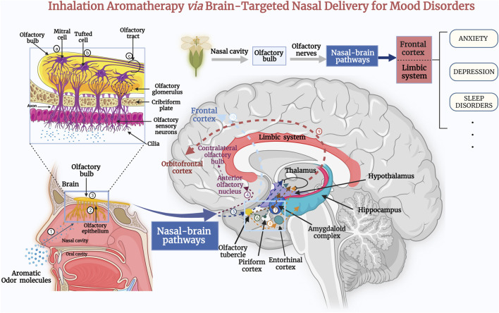
FIGURE 1.
Inhalation of the extracts of aromatic plant via the nose sends signals directly to the olfactory system, where odor molecules target therapeutic drugs to brain tissue via nasal–brain channels. Subsequently, they act on the cerebral cortex, the thalamus, and the limbic system of the brain, and stimulate the brain to produce neurotransmitters to treat the symptoms of anxiety and depression, as well as improve sleep quality (Lv et al., 2013). The aromatic odor molecules are inhaled through the nasal cavity (1) to reach the olfactory epithelium (2) of the nasal mucosa (Schneider et al., 2018). First-order neurons transmit the odor-evoked response to the olfactory bulb (3). In the olfactory bulb, the axons of mitral cells (a) and some tufted cells (b) (secondary neurons) form the olfactory tract (c). The axons of some mitral cells or lateral branches enter the anterior olfactory nucleus (4) and pass to the contralateral olfactory bulb (Cha et al., 2021). Additional secondary neurons enter the olfactory striatum (medial, lateral, and medial) and then project to central olfactory areas, including the olfactory tubercle (5), piriform cortex (6), amygdala (7), and the entorhinal cortex (8). The entorhinal cortex partially transmits to the hippocampus. Eventually, the central olfactory-area signals are transmitted through the thalamus to the orbitofrontal cortex (9) (Lie et al., 2021). An additional olfactory signaling pathway passes directly from the central olfactory area to the prefrontal cortex (10). These impulses induce the release of neurotransmitters such as serotonin or endorphin, which act as a “bridge” between nerves and other bodily systems (Schneider et al., 2018; Smith and Bhatnagar, 2019).

