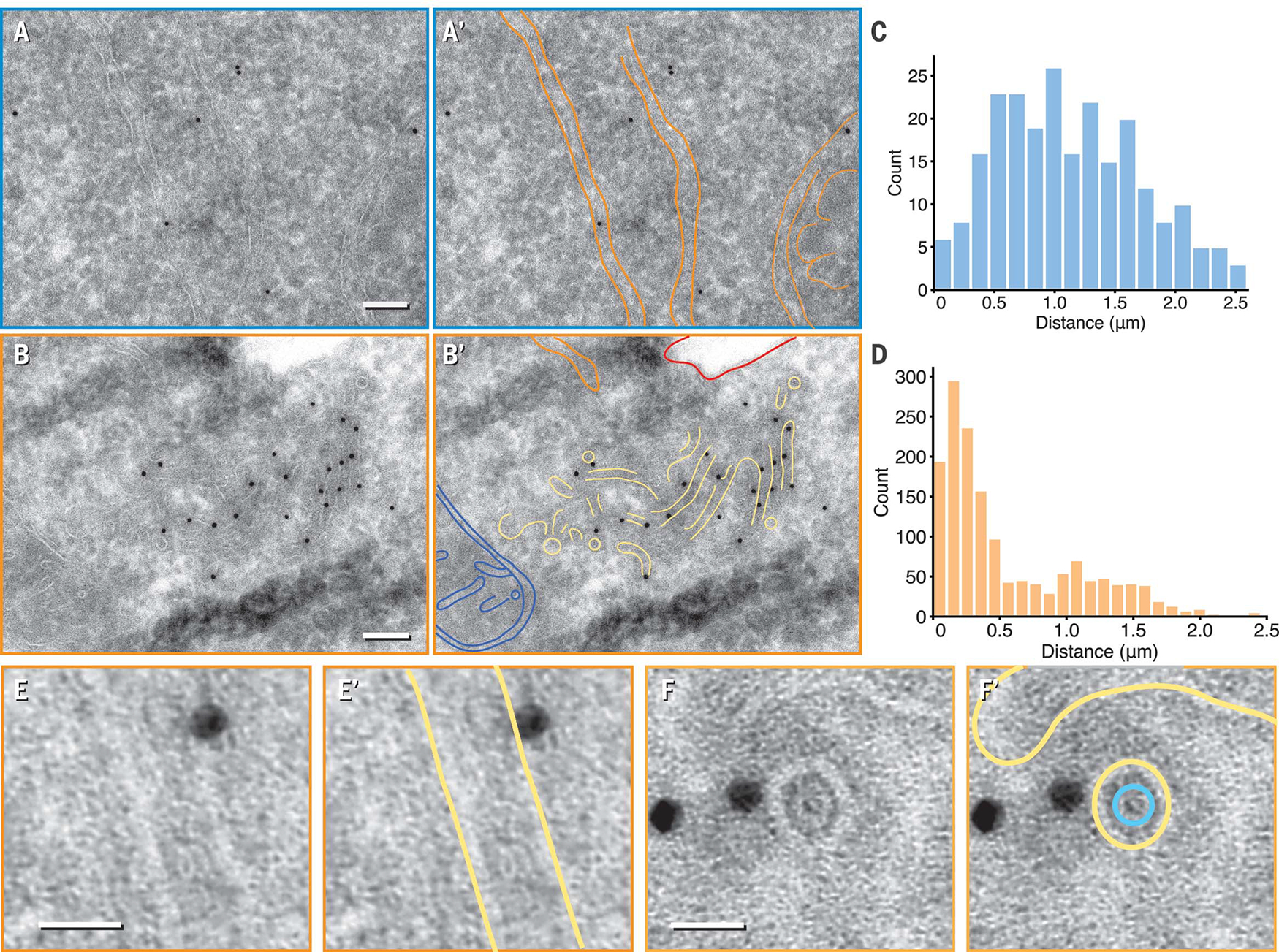
Fig. 2. Orthogonal immuno–electron microscopy revealed IRE1α subdomains.

(A and B) Representative micrograph of nonstressed (A) and stressed (B) HEK293-IRE1α-GFP. Gold particles recognizing the IRE1α-GFP epitope sparsely labeled general ER structures in nonstressed cells but localized to a region enriched in narrow membranes of 26 ± 2 nm in diameter (±SD) with stress. Orange, ER sheet and tubule membranes; blue, mitochondrion; red, plasma membrane; yellow, narrow membrane tubes. Scale bars are 100nm. (C and D) Histograms of inter–gold particle distances measured in micrographs from nonstressed (C) and stressed samples (D) revealed densely clustered gold particles enriched with ER-stress induction. (E to F) Enlarged longitudinal [(E) and (E′)] and end-on [(F) and (F′)] cross sections of ~28-nm diameter tubes that are close to gold particles. A ring-like density within the lumenal space was visible in (F′) (segmented in teal). Scale bars are 20 nm. Yellow, membrane.
