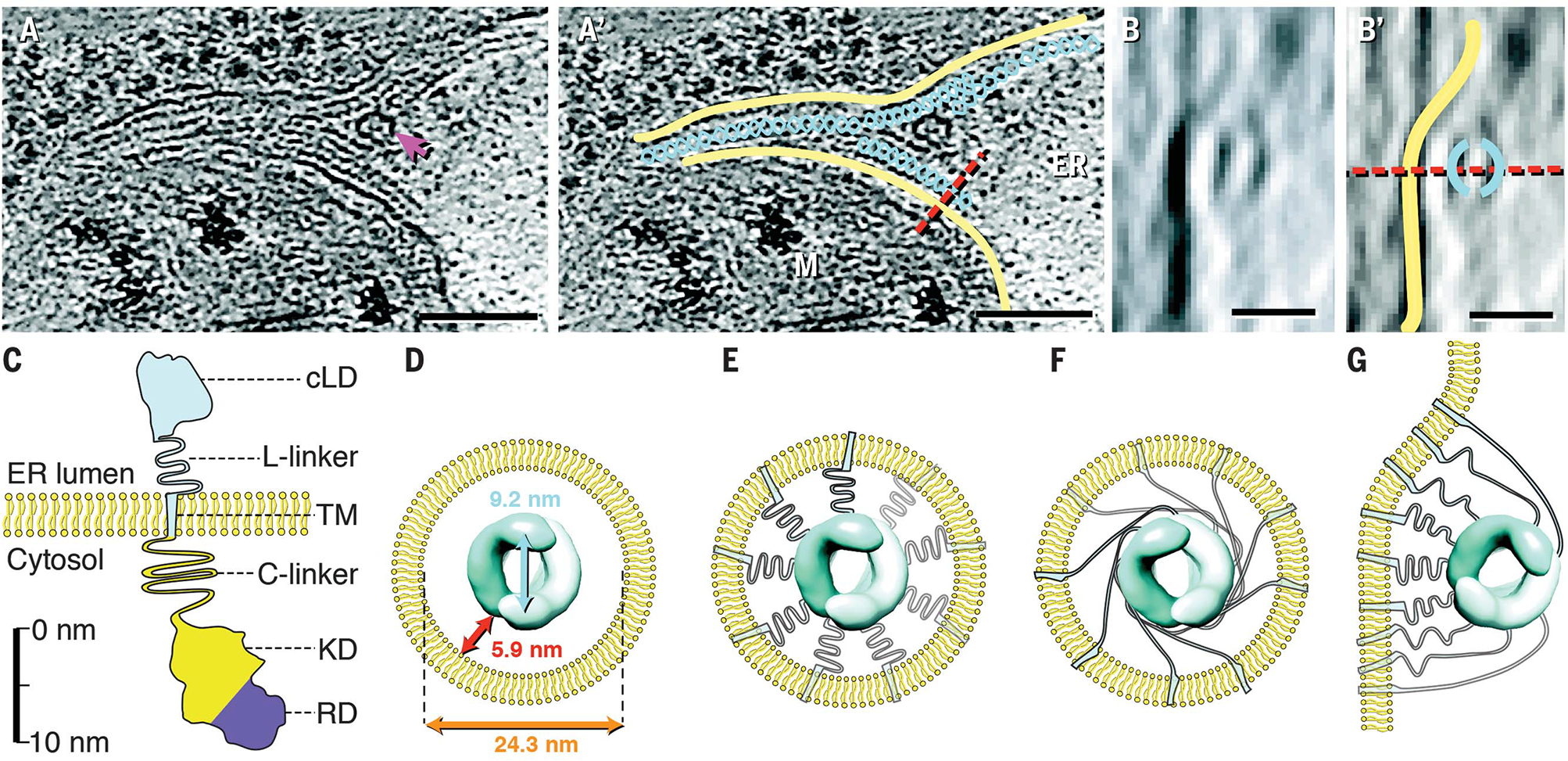
Fig. 5. IRE1α -LD helices accommodate a range of distances from membrane.

(A and A′) Instance of IRE1α-LD helices not enclosed by membrane tubes. The magenta arrow in (A) indicates a membrane fenestration. Yellow, membrane; teal, helical filaments; red dashed line, plane of rotation; ER, lumenal space; M, mitochondrion. Scale bars are 100 nm. (B and B′) Side view with 90° x-axis rotation along the indicated plane. Color coding is the same as in (A′). Scale bars are 20 nm. (C) Diagram of IRE1α domains drawn to approximate scale. cLD, core LD (amino acids 19 to 390); L-linker, lumenal linker (amino acids 391 to 443); TM, transmembrane helix (amino acids 444 to 464); C-linker, cytoplasmic linker (amino acids 465 to 570); KD, kinase domain (amino acids 571 to 832); RD, RNase domain (amino acids 835 to 964). (D) Dimensions of IRE1α-LD helices within IRE1α subdomain lumenal space. (E and F) Schematics for alternative TM and L-linker arrangements. There were 18 monomers per turn per double helix but only nine TMs and L-linkers were shown for clarity. (G) Model for TM and L-linker arrangement for helices as seen in (B).
