FIG. 3.
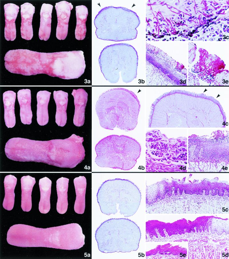
Fig. 3. Oral candidiasis in immunosuppressed rats; untreated control group. (a and b) Macroscopic (a) and panoramic (b) histological lingual candidiasis; the epithelium of the tongue shows irregular thickness and hyperkeratotic areas with plentiful hyphae (arrowheads). PAS; magnification, ×5. (c) Diffuse, abundant Candida mycelial elements affecting the superficial layers of the epithelium. PAS; magnification, ×600. (d) Large focal group of hyphae penetrating into the deeper layers of the epithelium. PAS; magnification, ×125. (e) Magnification of panel d. Note the abundant hyphae that have destroyed the keratinocytes, associated with small leukocyte infiltrates. PAS; magnification, ×600. Fig. 4. Oral candidiasis in immunosuppressed rats; animals treated with GM237354 at 1.25 mg/kg TID for 7 days. (a and b) Macroscopic (a) and panoramic (b) histological lingual candidiasis; the transverse sections show important focal thickening of the epithelium (arrowhead). HE; magnification, ×5. (c) Multifocal hyperplasia of the epithelium, associated with irregular accumulations of hyphae (arrowheads). PAS; magnification, ×15. (d) Intraepithelial microabscess containing numerous leukocytes, neutrophils, and partially degenerated hyphae. PAS; magnification, ×600. (e) Destruction of several epithelial layers, with extensive neutrophil infiltration in relation to the presence of hyphae in the epithelium. PAS; magnification, ×125. Fig. 5. Oral candidiasis in immunosuppressed rats; animals treated with GM237354 at 10 mg/kg TID for 7 days. (a and b) Macroscopic (a) and panoramic (b) histological lingual candidiasis; note the different coloration of the dorsal surface of the tongue, due to the alternation of hyperplastic and normal epithelial areas. PAS; magnification, ×5. (c) Basal cell hyperplasia, with scant differentiation of squamous cells and horny layer dyskeratosis. There are no C. albicans hyphae in the epithelial surface layer. PAS; magnification, ×125. (d) Focal alteration of the epithelial maturation of the tongue, reflected by intense dyskeratosis alternation associated with normal epithelium. A small increase of lymphocytes in the lamina propria was observed. HE; magnification, ×125. (e) Atrophy of the epithelium associated with intense regenerative changes. HE; magnification, ×125.

