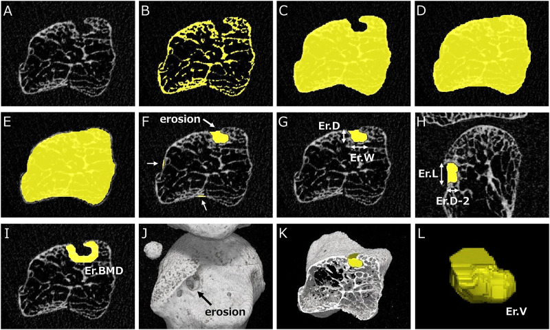
Fig 1. Algorithm for image analysis of erosion parameters for metacarpophalangeal (MCP) joints.
A. Gray-scale image of the metacarpal head in the axial view. B. Binarization at a threshold value of 320 mg/cm3. C. Filling bone marrow spaces. D. Filling concavities on bone surface. E. Shrinking the contour from Fig 1D by 0.25 mm. F. Extraction of all concavities (white arrows) and detection of erosions based on the false-positive pattern in Fig 2 and the definition from SPECTRA. G. Manual measurement of erosion depth (Er.D) and width (Er.W) in the axial view. H. Manual measurement of erosion depth (Er.D-2) and length (Er.L) in the perpendicular (coronal) view. I. Measurement of peripheral BMD (Er.BMD) surrounding an erosion at a width of 1 mm. J. 3D image of MCH and erosion on the radial side (black arrow). K. Axial section of the 3D image including an erosion. L. Volume of interest (VOI) of an erosion and measurement of erosion volume (Er.V).

