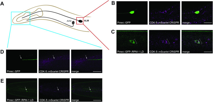
Fig 6. CDK-5 colocalizes with RPM-1 LD in neuronal soma.
A) Schematic showing ALM mechanosensory neuron and region imaged for axon termination site (blue) and soma (red). B) CDK-5::mScarlet CRISPR is present in the soma of ALM neurons (bracket) visualized using transgenic GFP expressed in mechanosensory neurons (muIs32). C) CDK-5::mScarlet colocalizes with GFP::RPM-1 LD in ALM soma (bracket). D) Representative images showing CDK-5::mScarlet is not detected at the ALM axon tip visualized using transgenic GFP (arrow). E) GFP::RPM-1 LD accumulates at the ALM axon tip (arrow) where CDK-5::mScarlet is not detected. Scale bars 10 μm.

