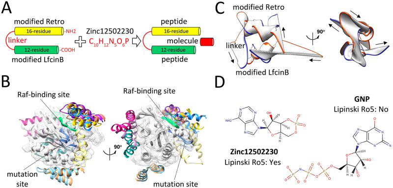
Fig 5. Binding energy and structural evaluations of final selected peptides and molecule.
(A) Schematic representation of dimer peptide construction and combination of this peptide with Zinc12502230. (B) Ten top-ranked peptide-protein complexes obtained from flexible docking of the modified Retro-LfcinB peptide (each pose in a different color) with K-RasG12D (gray). The Raf-binding and mutation sites are shown in green and blue colors, respectively. (C) The dominant mode of motion resulted from 50 frames of Retro-LfcinB MD simulations. The orange and blue colors represent the first and last frames, respectively. Arrows show the direction of motions. (D) Molecular representation of the small molecule Zinc12502230 (left) and GNP (right).

