
Fig. 5.
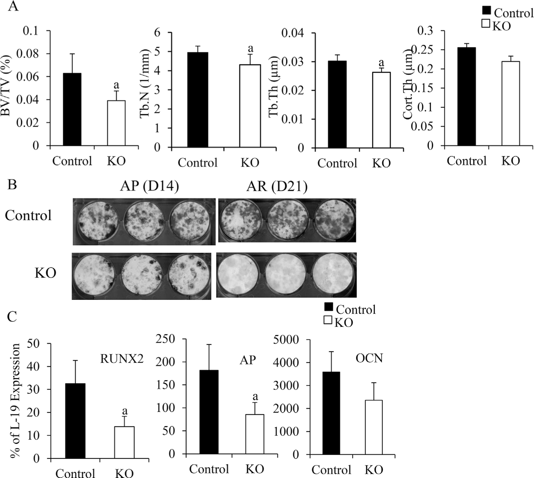
The effects of deletion of IGF-IR in the osterix-expressing cells on bone. At 7 weeks, bone structure of the KO (open bars) and control (solid bars) were determined by μCT (A), n = 4 in control group, n = 5 in KO group. Osteoblast differentiation (B) was examined by bone marrow stromal cell culture at D14 (AP staining) and D21 (alizarin red [AR] staining of mineralized nodules). RNA was isolated from the bones (marrow flushed out). mRNA levels of osteoblast differentiation markers RUNX2, AP, and osteocalcin (OCN) were measured by QPCR (C). In A and C, results are expressed by means SD. ± a: p < 0.05 KO versus control.