. 2022 Apr 26;11:e74275. doi: 10.7554/eLife.74275

Figure 5. Inhibitory proteins do not prevent Pro-σK(1–127) from interacting with SpoIVFB.

(A) Pro-σK(1–127), SpoIVFA, and GFPΔ27BofA co-purify with cytTM-SpoIVFB. pET Quartet plasmids were used to produce a catalytically inactive E44C cytTM-SpoIVFB variant with a FLAG2 tag (pSO73), or a variant lacking FLAG2 as a negative control (pSO149), in combination with Pro-σK(1–127), SpoIVFA, and GFPΔ27BofA in Escherichia coli. Samples collected after 2 hr of IPTG induction were subjected to co-immunoprecipitation with anti-FLAG antibody beads. Input, unbound, 1/10 bound (diluted to match input), and (undiluted) bound samples were subjected to immunoblot analysis with FLAG, penta-His, SpoIVFA, and GFP antibodies. The single star (*) indicates cross-reacting proteins below SpoIVFA and the double star (**) indicates a cross-reacting protein or breakdown species of GFPΔ27BofA that fail to co-purify. A representative result from two biological replicates is shown. (B) A231C in the cytTM-SpoIVFB CBS domain forms disulfide cross-links with A97C in the C-terminal region of Pro-σK(1–127) in the absence or presence of inhibitory proteins. pET Duet plasmids were used to produce single-Cys A231C cytTM-SpoIVFB E44Q in combination with single-Cys A97C Pro-σK(1–127) (pSO130; squares and line with long dashes labeled ‘No BofA’, although SpoIVFA is also absent) or with Cys-less Pro-σK(1–127) as a negative control (pSO255; crosses and line labeled ‘Cys-less’) in E. coli. pET Quartet plasmids were used to produce single-Cys A231C cytTM-SpoIVFB E44Q, single-Cys A97C Pro-σK(1–127), and Cys-less SpoIVFA in combination with Cys-less MBPΔ27BofA (pSO133; circles and line labeled ‘MBPΔ27BofA’) or with Cys-less full-length BofA (pSO246; diamonds and line labeled ‘BofA’) in E. coli. Samples collected after 2 hr of IPTG induction were treated for 15, 30, 45, or 60 min with Cu2+(phenanthroline)3 oxidant to promote disulfide bond formation and subjected to immunoblot analysis with FLAG antibodies to visualize the cytTM-SpoIVFB monomer, dimer, and complex with Pro-σK(1–127) (Figure 5—figure supplement 3C). Abundance of the complex was divided by the total amount of cytTM-SpoIVFB monomer, dimer, and complex. The ratio over time was plotted (n=2) with a best-fit trend line.

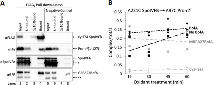
Figure 5—source data 1. Immunoblot images (raw and annotated) (Figure 5A) and quantification of cross-linking (Figure 5B).

Figure 5.

Figure 5—figure supplement 1. Neither GFPΔ27BofA nor full-length BofA when coproduced with SpoIVFA prevent Pro-σK(1–127) from interacting with SpoIVFB.

Figure 5—figure supplement 1.

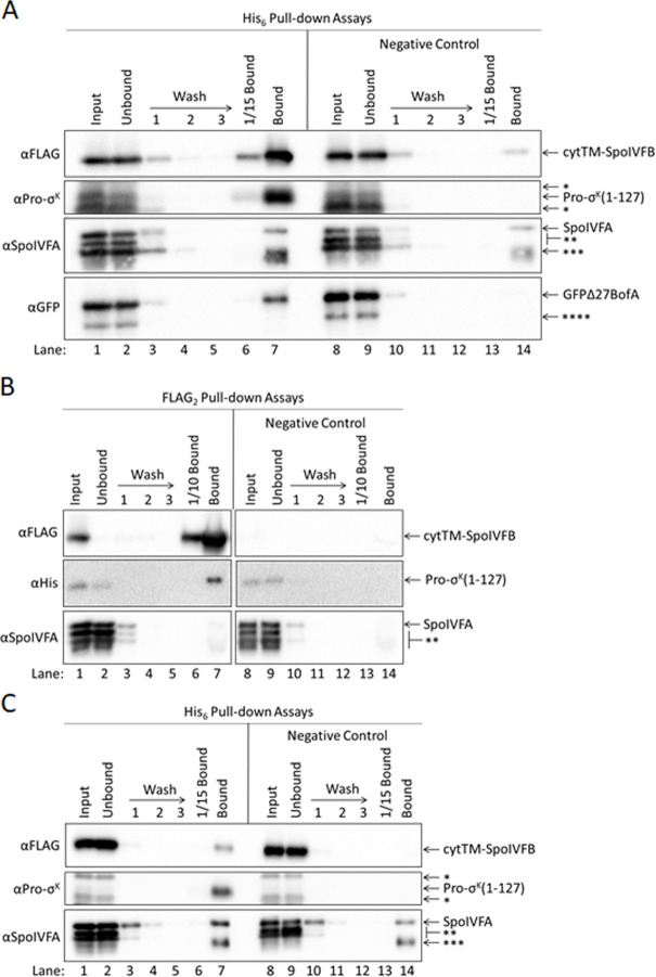
(A) GFPΔ27BofA and SpoIVFA do not prevent SpoIVFB from co-purifying with Pro-σK(1–127). pET Quartet plasmids were used to produce Pro-σK(1–127) (pSO73), or a variant lacking His6 as a negative control (pSO82), in combination with a catalytically inactive E44C cytTM-SpoIVFB variant containing FLAG2 but lacking His6, GFPΔ27BofA, and SpoIVFA in Escherichia coli. Samples collected after 2 hr of IPTG induction were subjected to co-purification with cobalt resin. Input, unbound, wash, 1/15 bound (diluted to match input), and (undiluted) bound samples were subjected to immunoblot analysis with FLAG, Pro-σK, SpoIVFA, and GFP antibodies as indicated. The single star (*) indicates cross-reacting proteins above and below Pro-σK(1–127) that fail to co-purify. The double star (**) indicates cross-reacting proteins below SpoIVFA that fail to co-purify. The triple star (***) indicates a putative breakdown species of SpoIVFA that appears to co-purify, but also binds nonspecifically. The quadruple star (****) indicates a cross-reacting protein or breakdown species of GFPΔ27BofA that fails to co-purify. All four proteins were seen in the bound sample (lane 7). Only Pro-σK(1–127) and the cytTM-SpoIVFB variant were detected in the diluted bound sample (lane 6). Most of the cytTM-SpoIVFB variant, GFPΔ27BofA, and SpoIVFA were observed in the unbound sample (lane 2), indicating inefficient co-purification. A negative control with a Pro-σK(1–127) variant lacking the His6 tag showed none of the Pro-σK(1–127) variant or GFPΔ27BofA in the bound sample, but a small amount of the cytTM-SpoIVFB variant and considerable SpoIVFA were detected (lane 14), indicative of nonspecific binding to the resin. In the case of SpoIVFA, nonspecific binding rather than co-purification with Pro-σK(1–127) appears to account for most of the signal in lane 7. A putative SpoIVFA breakdown species (indicated by ***) exhibited a similar pattern of abundance in samples as intact SpoIVFA. (B) Full-length BofA and SpoIVFA do not prevent Pro-σK(1–127) from co-purifying with SpoIVFB. pET Quartet plasmids were used to produce a catalytically inactive E44C cytTM-SpoIVFB variant containing FLAG2 but lacking His6 (pSO215), or a variant lacking FLAG2 as a negative control (pSO217), in combination with Pro-σK(1–127), BofA, and SpoIVFA in E. coli. Samples collected after 2 hr of IPTG induction were subjected to co-immunoprecipitation with anti-FLAG antibody beads. Input, unbound, wash, 1/10 bound (diluted to match input), and (undiluted) bound samples were subjected to immunoblot analysis with FLAG, penta-His, and SpoIVFA antibodies as indicated. Stars indicate proteins as in (A). (C) Full-length BofA and SpoIVFA do not prevent SpoIVFB from co-purifying with Pro-σK(1–127). pET Quartet plasmids were used to produce Pro-σK(1–127) (pSO215), or a variant lacking His6 as a negative control (pSO216), in combination with a catalytically inactive E44C cytTM-SpoIVFB variant containing FLAG2 but lacking His6, BofA, and SpoIVFA in E. coli. Samples collected after 2 hr of IPTG induction were subjected to co-purification with cobalt resin. Input, unbound, wash, 1/15 bound (diluted to match input), and (undiluted) bound samples were subjected to immunoblot analysis with FLAG, Pro-σK, and SpoIVFA antibodies as indicated. Stars indicate proteins as in (A). A representative result from two biological replicates is shown in each panel. BofA and SpoIVFA did not completely prevent Pro-σK(1–127) from interacting with the cytTM-SpoIVFB variant in (B) or (C) (lane 7 in each panel). We note that coproduction of BofA decreased the accumulation of Pro-σK(1–127) in the input samples (lane 1 in each panel) compared to coproduction of GFPΔ27BofA (lane 1 in Figure 5A and in (A)). We also note that SpoIVFA failed to co-purify with the cytTM-SpoIVFB variant when BofA was coproduced ((B), lane 7), in contrast to the result when GFPΔ27BofA was coproduced (Figure 5, lane 4). Perhaps BofA decreased Pro-σK(1–127) accumulation and SpoIVFA co-purification more than GFPΔ27BofA because TMS1 in full-length BofA hinders the interaction between Pro-σK(1–127) and the cytTM-SpoIVFB variant, making Pro-σK(1–127) more susceptible to degradation, which may impair SpoIVFA co-purification.
Figure 5—figure supplement 1—source data 1. Immunoblot images (raw and annotated).
Figure 5—figure supplement 2. GFPΔ27BofA and SpoIVFA do not prevent full-length Pro-σK from interacting with SpoIVFB.

Figure 5—figure supplement 2.

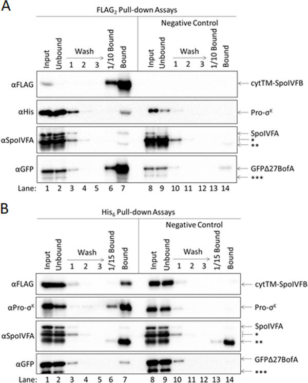
(A) GFPΔ27BofA and SpoIVFA do not prevent Pro-σK from co-purifying with SpoIVFB. pET Quartet plasmids were used to produce a catalytically inactive E44C cytTM-SpoIVFB variant containing FLAG2 but lacking His6 (pSO211), or a variant lacking FLAG2 as a negative control (pSO221), in combination with Pro-σK-His6, GFPΔ27BofA, and SpoIVFA in Escherichia coli. Samples collected after 2 hr of IPTG induction were subjected to co-immunoprecipitation with anti-FLAG antibody beads. Input, unbound, wash, 1/10 bound (diluted to match input), and (undiluted) bound samples were subjected to immunoblot analysis with FLAG, penta-His, SpoIVFA, and GFP antibodies as indicated. The single star (*) indicates cross-reacting proteins below SpoIVFA that fail to co-purify. The double star (**) indicates a putative breakdown species of SpoIVFA that appears to co-purify, but also binds nonspecifically. The triple star (***) indicates a cross-reacting protein or breakdown species of GFPΔ27BofA that fails to co-purify. (B) GFPΔ27BofA and SpoIVFA do not prevent SpoIVFB from co-purifying with Pro-σK. pET Quartet plasmids were used to produce Pro-σK-His6 (pSO211), or a variant lacking His6 as a negative control (pSO220), in combination with a catalytically inactive E44C cytTM-SpoIVFB variant containing FLAG2 but lacking His6, GFPΔ27BofA, and SpoIVFA in E. coli. Samples collected after 2 hr of IPTG induction were subjected to co-purification with cobalt resin. Input, unbound, wash, 1/15 bound (diluted to match input), and (undiluted) bound samples were subjected to immunoblot analysis with FLAG, Pro-σK, SpoIVFA, and GFP antibodies as indicated. Stars indicate proteins as in (A). A representative result from two biological replicates is shown in each panel. We note that when Pro-σK-His6 was coproduced rather than Pro-σK(1–127), less SpoIVFA and more GFPΔ27BofA co-purified with the cytTM-SpoIVFB variant (compare (A) and Figure 5A), and less of both inhibitory proteins co-purified with Pro-σK-His6 (compare (B) and Figure 5—figure supplement 1A), consistent with the notion that the C-terminal half of full-length Pro-σK affects complex formation.
Figure 5—figure supplement 2—source data 1. Immunoblot images (raw and annotated).
Figure 5—figure supplement 3. Disulfide cross-linking between the cytTM-SpoIVFB CBS domain and the Pro-σK(1–127) C-terminal region.

Figure 5—figure supplement 3.

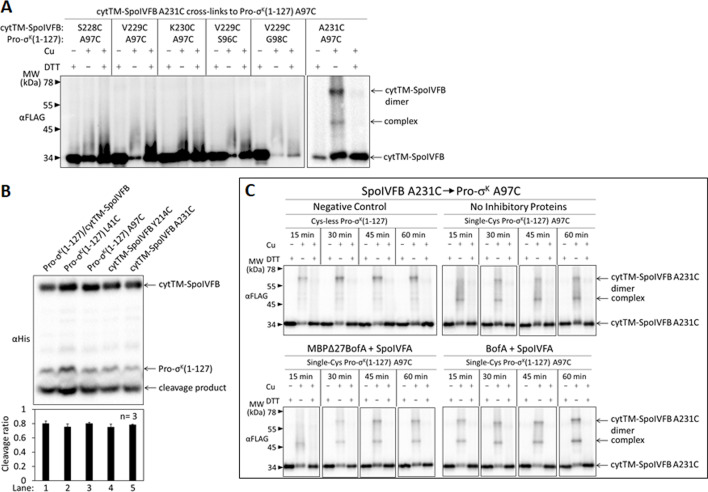
(A) Cross-linking between single-Cys cytTM-SpoIVFB variants and single-Cys Pro-σK(1–127) variants. pET Duet plasmids (pSO122–pSO126 and pSO130) were used to produce single-Cys S228C, V229C, K230C, or A231C cytTM-SpoIVFB E44Q variants in combination with single-Cys S96C, A97C, or G98C Pro-σK(1–127) variants in Escherichia coli. Samples collected after 2 hr of IPTG induction were treated and subjected to immunoblot analysis as explained in the Figure 4A legend. A representative result from two biological replicates is shown. (B) Cleavage assays examining the effects of Cys substitutions in cytTM-SpoIVFB or Pro-σK(1–127). pET Duet plasmids were used to produce Pro-σK(1–127) and cytTM-SpoIVFB from pYZ2 as a control (lane 1), cytTM-SpoIVFB and the indicated Cys-substituted Pro-σK(1–127) from pSO157 or pSO158 (lanes 2 and 3), or Pro-σK(1–127) and the indicated Cys-substituted cytTM-SpoIVFB from pSO159 or pSO160 (lanes 4 and 5) in E. coli. Samples collected after 2 hr of IPTG induction were subjected to immunoblot analysis, and the graph shows quantification of the cleavage ratio, as explained in the Figure 1B legend. (C) Time course of cross-linking between the single-Cys A231C CBS domain variant of cytTM-SpoIVFB E44Q and single-Cys A97C Pro-σK(1–127) in the absence or presence of inhibitory proteins. See the Figure 5B legend for explanation of the experiment. A representative result from two biological replicates is shown.
Figure 5—figure supplement 3—source data 1. Immunoblot images (raw and annotated) and quantification of cleavage assays (Figure 5—figure supplement 3B).