. 2022 Apr 26;11:e74275. doi: 10.7554/eLife.74275

Figure 7. Model of SpoIVFB monomer with BofA and parts of SpoIVFA and Pro-σK.

(A) Model of SpoIVFB, BofA TMS2, and the C-terminal part of Pro-σK(1–127). At Left, a side view of the complex, showing the six TMSs of the SpoIVFB membrane domain with the active site zinc ion (magenta), the interdomain linker, and the CBS domain (green), BofA TMS2 (cyan), and Pro-σK(38–114) (red). In the enlarged view of the active site cleft (Center), TMSs 1–6 of SpoIVFB and TMS2 of BofA are numbered. At Right, a top view is shown. (B) Model of SpoIVFB with full-length BofA and Pro-σK(38–114). Predicted TMSs 1 and 2 of BofA are numbered and its C-terminal region is labeled ‘C’ near the C-terminus in the views shown in the Center and at Right. (C) Model of SpoIVFB with full-length BofA, SpoIVFA(65–111) (purple, residue 111 is numbered), and Pro-σK(38–114). TMS, transmembrane segment.

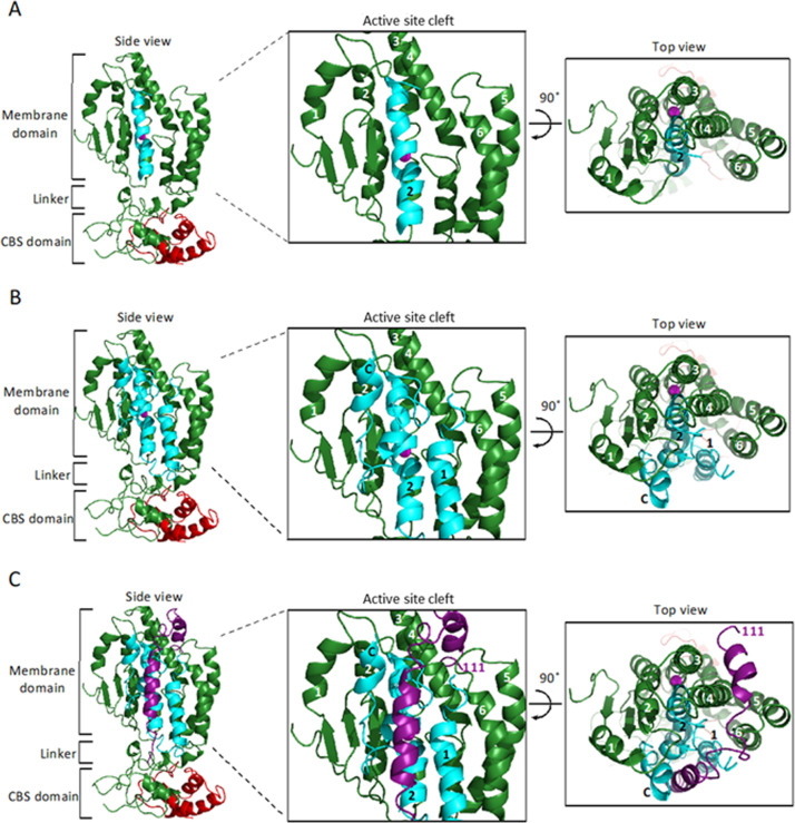
Figure 7—source data 1. PyMOL session file used to produce the images and PDB file of the model of a SpoIVFB tetramer with BofA and parts of SpoIVFA and Pro-σK.

Figure 7.

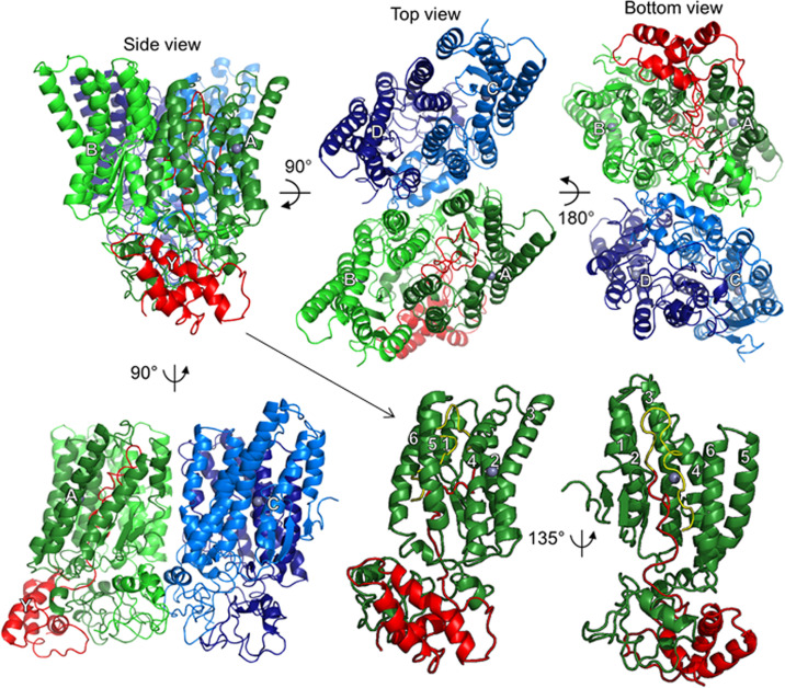
Figure 7—figure supplement 1. Model of SpoIVFB tetramer with part of one Pro-σK molecule.

Figure 7—figure supplement 1.

At Upper Left, a side view shows the SpoIVFB membrane domains at the top and the CBS domains at the bottom, facing the A (dark green) and B chains (light green), whose CBS domains primarily provide the dimerization interface. The SpoIVFB A chain also interacts with the Pro-σK Y chain (residues 1–114) (red). The top view (Center) reveals the SpoIVFB C (light blue) and D (dark blue) chains, whose CBS domains dimerize. The bottom view (Upper Right) emphasizes the CBS domains, as well as the interface between the A/B and C/D dimers, formed primarily by the CBS domains of the B and C chains. At Lower Left, a side view facing the A and C chains also shows the interface between the A/B and C/D dimers of the SpoIVFB tetramer. Each SpoIVFB chain is labeled near its zinc ion (gray), which is hidden in some views. At Lower Center, a side view of the SpoIVFB A chain monomer (TMSs 1–6 labeled) interacting with the Pro-σK Y chain (Proregion residues 1–21 yellow and σK residues 22–114 red), in the same orientation as at Upper Left (hence the arrow), but with the other SpoIVFB chains hidden. At Lower Right, a side view into the active site cleft of the SpoIVFB A chain emphasizes proximity between the zinc ion and the cleavage site in the Pro-σK Y chain (between residues 21 and 22). TMS, transmembrane segment.
Figure 7—figure supplement 1—source data 1. PyMOL session file used to produce the images and PDB file of the model of a SpoIVFB tetramer with part of one molecule of Pro-σK.
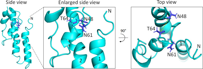
Figure 7—figure supplement 2. Model of BofA showing conserved residues important for SpoIVFB inhibition.

Figure 7—figure supplement 2.

At Left, a side view of full-length BofA in the same orientation as Figure 7C shows the side chains of conserved residues N48, N61, and T64 colored blue. Numbers indicate predicted TMSs 1 and 2, and N indicate the N-terminus. In the enlarged side view (Center), labels indicate the conserved residues important for SpoIVFB inhibition. At Right, a top view shows the 5.6 Å distance (dashed yellow line) between the Cβ atoms of the N48 and T64 side chains that may interact. The model predicts that the N61 side chain points away from the N48 and T64 side chains, but the orientation of the N61 and T64 side chains is uncertain given the predicted loop structure. TMS, transmembrane segment.
Figure 7—figure supplement 2—source data 1. PyMOL session file used to produce the images.