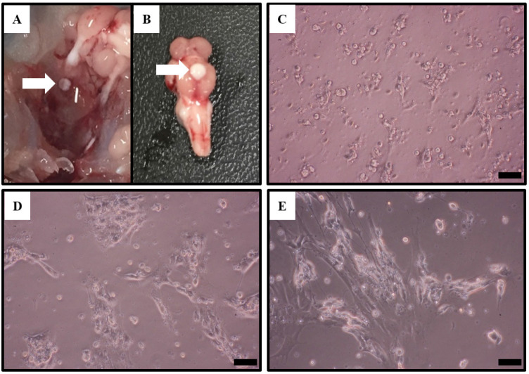
Fig. 1. Photographs of Nile tilapia pituitary and microscopic observation of pituitary cell culture.
(A) Location and isolation of Nile tilapia pituitary (arrow). (B) Location of the pituitary in Nile tilapia. (C–E) The pituitary of Nile tilapia was cultured for 24 (C), 48 (D) and 72 (E) h and then photographed by a camera. Scale bar=100 μm.

