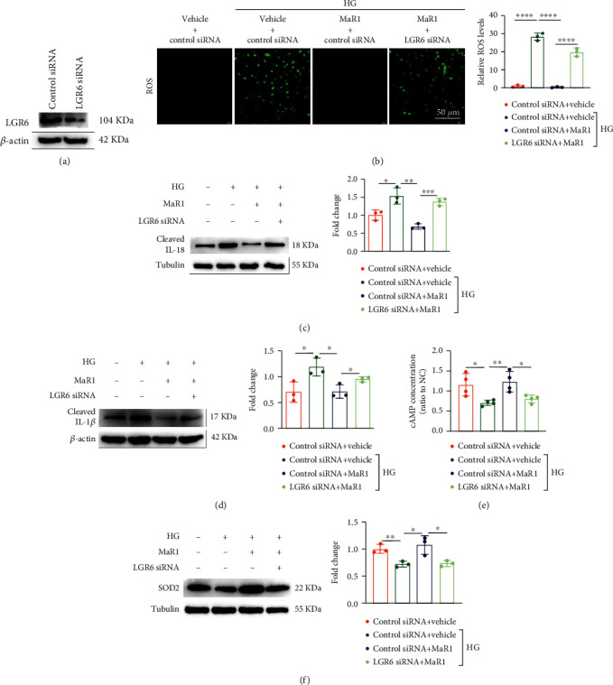
Figure 6.

MaR1 alleviated high glucose-induced inflammation via LGR6-mediated antioxidant pathway. (a) Western blotting analysis images showing the knockout efficiency of LGR6 siRNA. (b) DCFH-DA probe was used to detect the levels of ROS in high glucose-stimulated HK-2 cells of LGR6 knock-down with MaR1 intervention. Bar: 50 μm. (c) Western blotting analysis images showing the levels of IL-18 in cell culture supernatant of high glucose-stimulated HK-2 cells of LGR6 knock-down with MaR1 intervention. (d) Western blotting analysis images showing the levels of IL-1β in cell culture supernatant of high glucose-stimulated HK-2 cells of LGR6 knock-down with MaR1 intervention. (e) The levels of cAMP in high glucose-stimulated HK-2 cells of LGR6 knock-down with MaR1 intervention. (f) Representative western blotting analysis images showing the protein levels of SOD2 in high glucose-stimulated HK-2 cells of LGR6 knock-down with MaR1 intervention. All results are representative of three independent experiments. Data were expressed as mean ± SD; ∗P <0.05; ∗∗P <0.01; ∗∗∗P <0.001; ∗∗∗∗P <0.0001.
