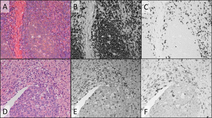
Fig. 4. H&E and dual IHC staining of tonsil for CD20 & CD8 (A-C) and colon tumor for CD3 and CD8 (D-F).
A and D Color camera images of transmitted visible light showing H&E staining. B, C Monochrome camera images of transmitted light at 405 nm (filtered tungsten lamp) where DCC CDC absorbs light, staining CD20 (B), and 769 nm (filtered tungsten lamp) where Cy7 CDC absorbs light, staining CD8 (C). Monochrome camera images of transmitted light at 385 nm (LED) where HCC CDC absorbs light, staining CD3 (E), and 770 nm (LED) where Cy7 CDC absorbs light, staining CD8 (F). Images were recorded using a ×20 objective.

