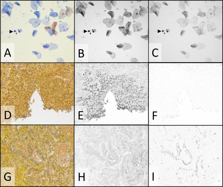
Fig. 5. Cytological stain and special stain plus duplex IHC: PAP-staining of cervical cytology specimen (A–C) and mucicarmine special staining of NSCLC SCC (D–F) and ADC (G–I).
Color camera images of transmitted visible light showing PAP (A) and mucicarmine special stain (D & G) staining. Monochrome camera images of transmitted light at 405 nm and 770 nm where DCC (B) and Cy7 (C) absorb light, staining Ki-67 (B) and p16 (C). Monochrome camera images of transmitted light at 769 nm where Cy7 CDC absorbs light, staining p40 (E and H), and 880 nm where ir870 CDC absorbs light, staining TTF-1 (F and I). Images were recorded using a ×20 objective.

