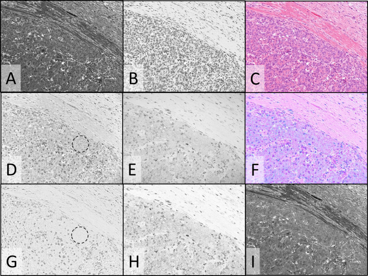
Fig. 6. Multispectral imaging and image processing: composite color images and spectral unmixing in colon tumor FFPE tissue stained with CD3/CD8 duplex IHC and H&E.
Monochrome images of transmitted light at 513, 620, 390, and 770 nm where eosin (A), hematoxylin (B), HCC CDC (D), and Cy7 (E) primarily absorb light, respectively, staining the two components of the H&E stain (A & B), CD3 (D) and CD8 (E). Spectrally unmixed images of CD3 (G), CD8 (H), and eosin (I). C Color composite image formed from the eosin (A) and hematoxylin (B) monochrome images. F Color composite image formed from the CD3 image (D), pseudo-colored magenta, and the CD8 image (E), pseudo-colored cyan. Dashed circles in (D) and (G) emphasize regions with faint hematoxylin crosstalk (D) that is removed after unmixing (G). Images were recorded using a ×20 objective.

