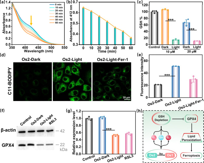
Fig. 5. Ferroptosis induction by Os2 under light irradiation.
a Irradiation time-dependent GSH (200 μM) depletion by Os2 (20 μM) upon blue light irradiation at 298 K and b the absorption at 412 nm was decreased by increasing the irradiation time. c The GSH levels in cells after different treatments. All the experiments were performed as duplicates of triplicates (n = 3 biologically independent samples, p values (p): 10 μM-0.000025, 20 μM-0.00065). d The fluorescence images of lipid peroxides in the treated cells detected by C11-BODIPY probe (30 μM, 310 K, 0.5 h). C11-BODIPY: λex = 488 nm, λem = 570 ± 50 nm. e The average fluorescence intensities calculated from the images in d. The experiment was repeated three times independently with similar results, p = 0.000024. f Western blot analysis of GPX4 in HeLa cells after treatment with Os2 (20 μM, 310 K, 8 h) with or without light treatment. RSL3 is the positive control group. g The relative expression levels of GPX4 calculated from f All the experiments were performed as duplicates of triplicates (n = 3 biologically independent samples, p = 0.000025). Error bars represent SD from the mean. Statistical significance was calculated with two-tailed Student’s t test (*p < 0.05, **p ≤ 0.01 or ***p ≤ 0.001). h The process of ferroptosis in this photoactive antitumor therapy. Light irradiation: 465 nm, 13 mW/cm2. GSH glutathione, Fer-1 ferrostatin-1, GPX4 glutathione peroxidase 4.

