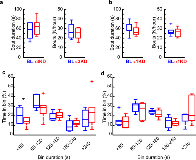
Fig. 2. α3KD, but not α1KD, in PV + TRN neurons decreased time spent in the shortest (<60 s) NREM bouts.
a Compared with their baseline conditions (BL; blue), α3KD (red) mice (n = 6) did not have altered durations or number of NREM bouts. b α1KD mice (n = 7) also did not have altered durations or number of NREM bouts. c The proportion of time spent in short bouts lasting <60 s was significantly reduced in α3KD mice (two-tailed paired t-test, *p = 0.03), but no other bins showed any differences between BL and α3KD (n = 6). d The proportion of time spend in bouts was unchanged in α1KD mice (n = 7). Box plots: center lines represent median values, 75th percentiles are box tops and 25th percentiles are box bottoms. + symbols represent outliers defined as >1.5x the interquartile range away from box tops or bottoms. Data from whole 12 h light period.

