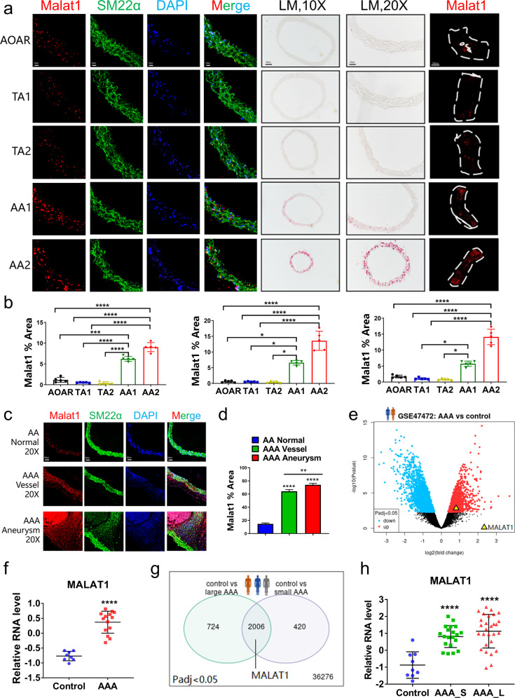
Fig. 5.
Detection of specific marker Malat1 of Sca1−VSMC 4 in different healthy aortic portions and AAA tissue. a Malat1, detected by fluorescence in situ hybridization (FISH) or whole-mount in situ hybridization (WISH), at both the transverse section and tissue fragments verified the distribution of VSMC 4. Scale bars = 10 μm, 50 μm, 20 μm, and 1000 μm, respectively. b The percentage of the Malat1-positive area in the AA portion either AA1 or AA2, was significantly higher than that in the others. n = 5 for each group. c Detection of the expression level of Malat1 in AAA, including both vessel and aneurysm parts, compared with that in the AA segment at baseline. Scale bar = 20 μm. d The percentage of the Malat1-positive area in the aneurysm was much higher than that in the normal abdominal aorta. n = 5 for each group. e Analysis of the human AAA database 1 (GSE47472, n = 14 patients in the AAA group, n = 8 donors in the control group) showed that MALAT1 was upregulated in human AAA tissue. f The relative RNA expression level was compared between AAA and control groups, in which MALAT1 was upregulated in the AAA group with a significant difference. g An analysis of human AAA database 1 (GSE57691, n = 28 patients in large AAA group, n = 20 patients in small AAA group, n = 9 donors in control group) provided evidence of differential expression of MALAT1. h The relative RNA expression level in AAA_L and AAA_S was significantly higher than that in the control group. AAA_L, large AAA. AAA_S, small AAA. Data were presented as mean ± SEM and normal distributions were tested by the Shapiro–Wilk method, which showed that all data were normally distributed. One-way ANOVA followed by Tukey post hoc test was used for (b, d, h). The student’s t test was used for (f). *P < 0.05, **P < 0.01, ***P < 0.001, ****P < 0.0001. Each experiment was repeated independently for a minimum of three times

