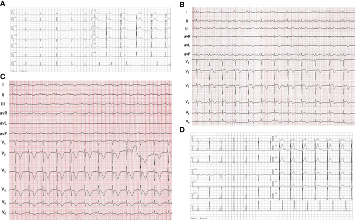
Figure 1.
Electrocardiograms. (A) Electrocardiogram obtained one month before this admission showed nearly normal. (B) Electrocardiogram at hospital admission showed QT prolongation and T-wave inversions in leads I and V1 to V6. (C) Electrocardiogram on the third hospital day showed more obvious QT prolongation and T-wave inversions in the limb leads (I, II, III, AVL, AVR and AVF) and the chest leads (V1 to V6). (D) Electrocardiogram after surgery showed QT prolongation and T-wave inversions disappeared.

