FIGURE 4.

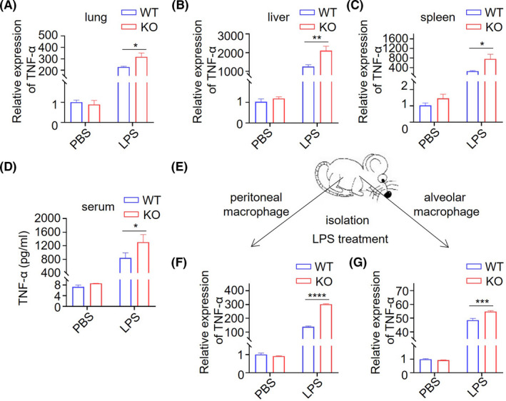
Trdmt1 deletion leads to impaired inflammatory factor response after LPS treatment. A‐C, The relative expression of TNF‐α in lung (A), liver (B), and spleen (C) of 1 mg/kg LPS‐treated Trdmt1 knockout rats. D, The TNF‐α protein level in serum of 1 mg/kg LPS‐treated Trdmt1 knockout rats. E‐H, Alveolar macrophage and peritoneal macrophage derived from Trdmt1 knockout rats were cultured and treated with 100 ng/mL LPS. The relative expression of TNF‐α of peritoneal macrophage (F), and alveolar macrophage (G) was determined. WT, wild‐type rats; KO, Trdmt1 knockout rats. Mean ± SEM; *P < 0.05, ***P < 0.001, ****P < 0.0001
