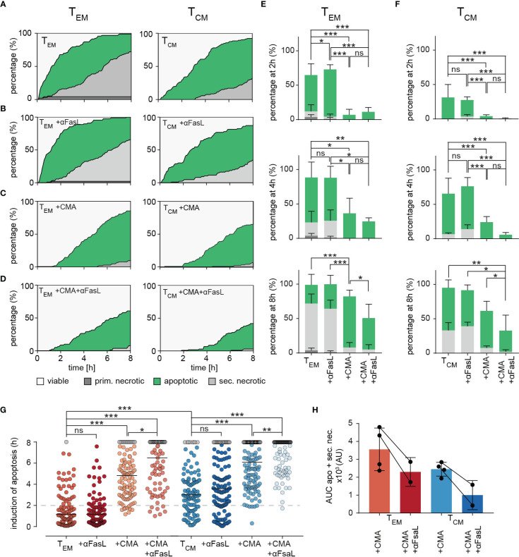
Figure 6.
Dissecting killing mechanisms of TEM and TCM. (A) To analyze target cell lysis by perforin-mediated or FasR-mediated killing mechanisms, sorted TEM or TCM and target cell were either un-treated as control (A) or treated with inhibiting FasL antibodies NOK-1 and NOK-2 (10 µg/each) (B), with 50 nM CMA (C) and with inhibiting FasL antibodies and CMA in combination (D). Effects of treatments on TEM and TCM are shown as representative death plots, respectively (A-D). (E, F) Quantification of target cell lysis at 2h, 4h and 8h for TEM (E) or TCM (F). (G) Δt of apoptosis for each analyzed target cell lysed by treated TEM or TCM in comparison to untreated (TEM or TCM) control cells. (H) Impact of FasL blocking quantified by analysis of area under the curve (AUC). Each point reflects the Δt of apoptosis induction from a single target cell. n=4 donors, TEM 94 cells, +αFasL 89 cells, +CMA 103, +CMA+αFasL (n=2 donors) 79, TCM 105 cells, +αFasL 122 cells, +CMA (n=2 donors) 122 cells, +CMA+αFasL 102 cells. Statistical analysis was done using a two-way ANOVA. *p<0.05; **p<0.01; ***p<0.001; ns, no significant difference.

