
FIG. 1.
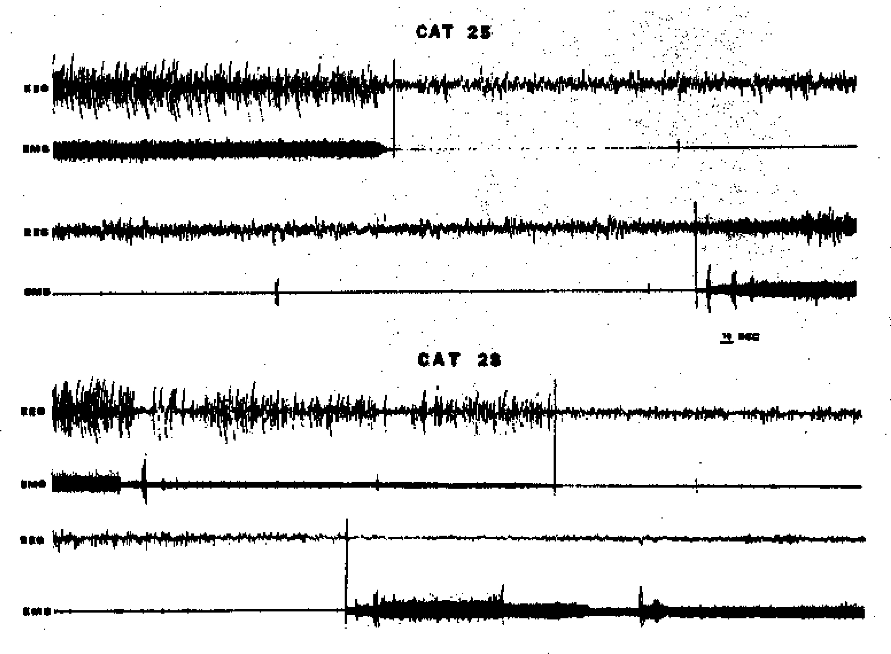
Four samples of the EEG and EMG data used in scoring for sleep state. The top tracings in each pair show a SWS-REM sleep transition. Note the EMG and EEG suppression at REM sleep onset. Cortical PGO spikes are clearly visible as negative monophasic deflections (downward) during REM sleep in Cat 28. The lower tracing in each pair shows a REM sleep offset. Note disappearance of PGO activity and return of muscle tone. Scoring judgments of REM sleep onset and offset are indicated by vertical lines across tracings.