
Fig. 5.
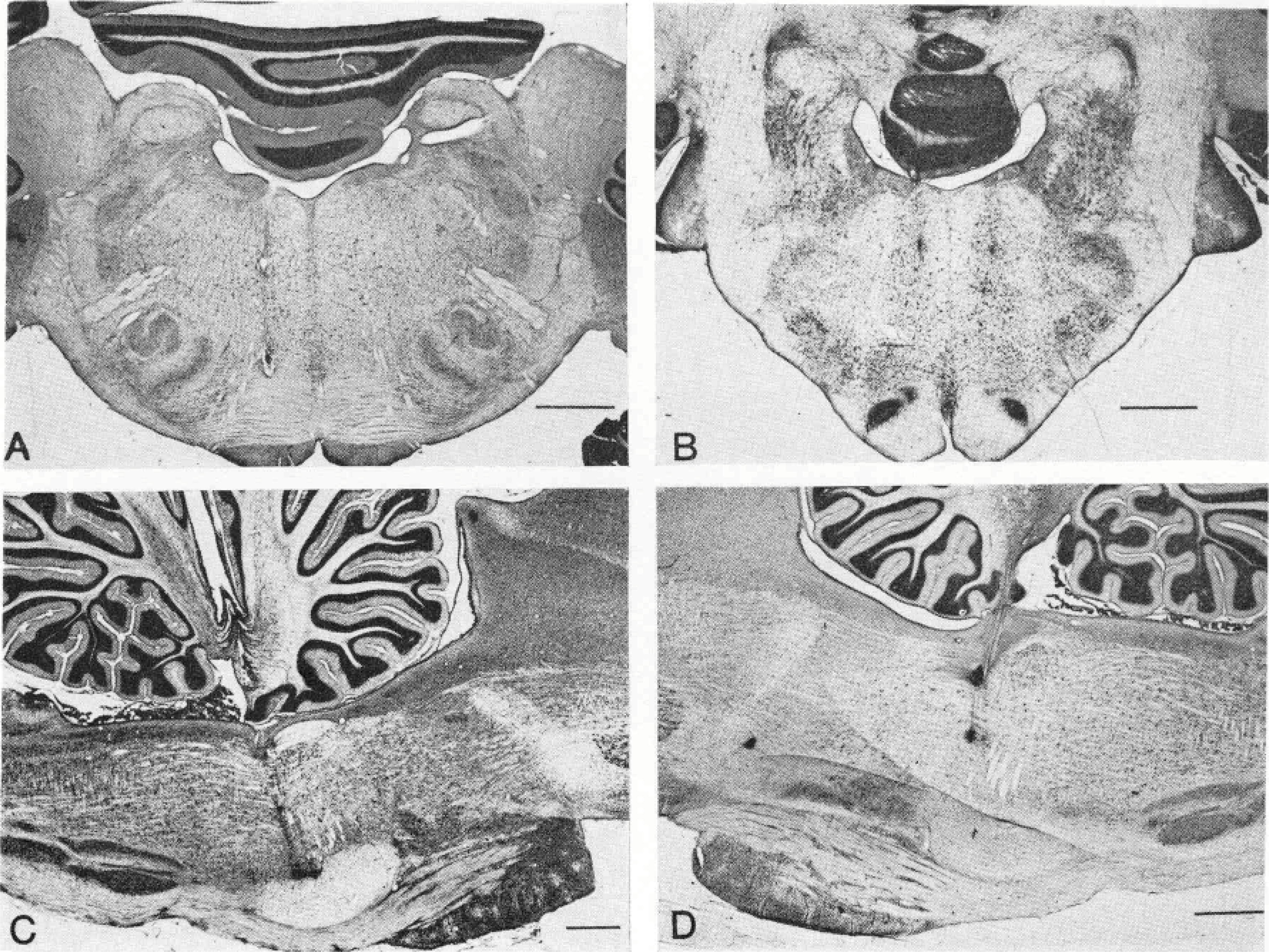
Representative electrode tracks. A—Coronal section at PS.2. Two electrode tracks are visible on left portion of slide. B—Coronal section at P7.7. A bilateral pair of electrode tracks is visible. C—Sagittal section at L1.2. A pair of electrode tracks and their terminal lesions are visible. D—Sagittal section at L1.2. An electrode track with two lesions. Calibration lines, 2 mm.