
Fig. 4.
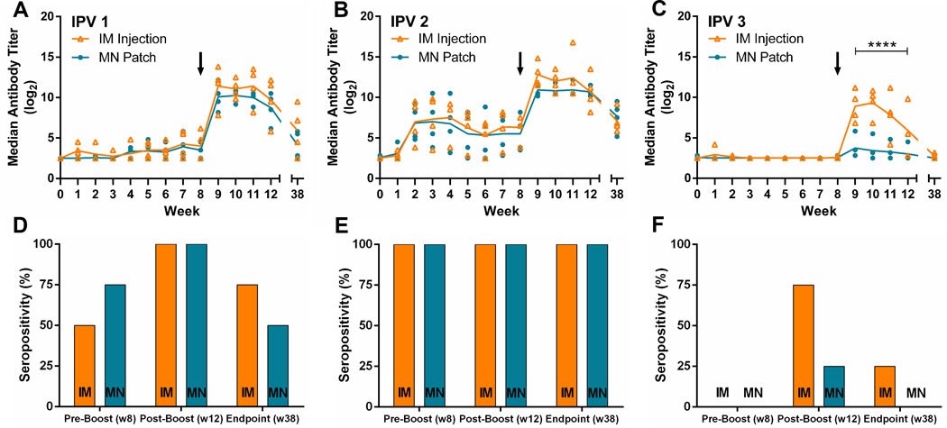
Serologic response and neutralizing antibody titers to poliovirus following vaccination. Rhesus macaques were vaccinated at week 0 and week 8 with IPV given either by microneedle (MN) patch or intramuscular (IM) injection. Serum was collected weekly and analyzed using a serotype-specific micro-neutralization assay, for IPV (A) type 1, (B) type 2 and (C) type 3. Seropositivity is also shown at weeks 8, 12, and 38 (D–F). Each data point represents a single animal while the lines represent the median of each group. The asterisks (****) represent a statistically significant difference (p < 0.0005) between the microneedle patch and intramuscular injection groups as measured by two-way ANOVA. Seropositivity was defined as a titer greater than or equal to 3.0 log2 [32,33].